2
Products based on this clone:
- {{heading}}
Please Select
- Ab00130-1.1 Anti-DC-SIGNR [16E7]
- Human
- Mouse IgG1
- Purified
- In Stock
- Ab00130-23.0 Anti-DC-SIGNR [16E7]
- Human
- Rabbit IgG
- Purified
- In Stock
Loading...
 United Kingdom (UK)
Recombinant monoclonal antibody to DC-SIGNR. Manufactured using AbAb’s Recombinant Platform with variable regions (i.e. specificity) from the hybridoma 16E7.
UniProt Accession Number of Target Protein: Q9H2X3
Alternative Name(s) of Target: CD299; C-type lectin domain family 4 member M; CD209 antigen-like protein 1; DC-SIGN-related protein; CLEC4M; Dendritic cell-specific ICAM-3-grabbing non-integrin 2; DC-SIGN2; Liver/lymph node-specific ICAM-3-grabbing non-integrin; L-SIGN
Immunogen: CD299
Specificity: Human CD299, which is an oligomeric type II transmembrane protein with a C-type lectin extracellular domain, the expression of which is restricted to immature DC, macrophages in the lung, and endothelial cells in the liver. It binds ICAM-3 and ICAM-7 to mediate the interaction of DC with T lymphocytes and endothelial cells in the initial stages of immune response and in the migratory behavior of DC. CD299 also binds the gp120 protein of HIV and the E2 envelope protein of HCV, thereby playing a role in viral infection.
Antibody first published in: PMID:
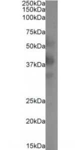
Western Blot using anti-DC-SIGNR (CLEC4M) antibody 16E7 (Ab00130) Jurkat cell extract (35µg protein in RIPA buffer) was resolved on a 10% SDS PAGE gel and blots probed with the chimeric rabbit version of 16E7 (Ab00130-23.0) at 0.1 µg/ml before detection using an anti-rabbit secondary antibody. A primary incubation of 1h was used and protein was detected by chemiluminescence. The expected band size for DC-SIGNR is 45.3kDa, though 9 other isoforms of this protein are known ranging in size from 22.4-44.7kDa (Uniprot ID: Q9H2X3). DC-SIGNR is also glycosylated at several positions. Ab00130-23.0 successfully detected the canonical human DC-SIGNR, as well as multiple other isoforms.
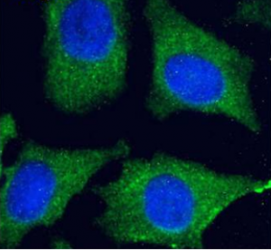
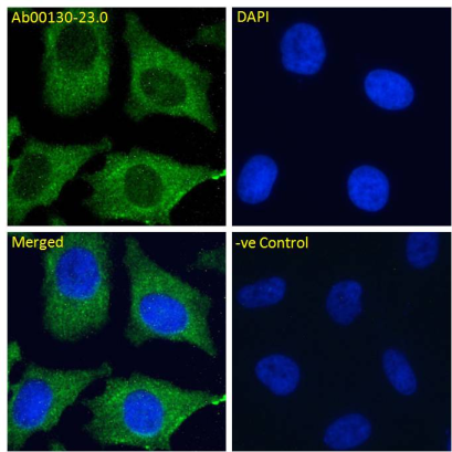
Immunofluoresence staining of fixed HeLa cells with anti-DC-SIGNR antibody 16E7 (Ab00130) Immunofluorescence analysis of paraformaldehyde fixed HeLa cells, permeabilized with 0.15% Triton stained with the chimeric rabbit IgG version of 16E7 (Ab00130-23.0) at 10 µg/ml for 1h followed by Alexa Fluor® 488 secondary antibody (1 µg/ml), showing cytoplasmic staining. The nuclear stain is DAPI (blue). Panels show from left-right, top-bottom Ab00130-23.0, DAPI, merged channels and a negative control. The negative control was stained with unimmunized rabbit IgG followed by Alexa Fluor® 488 secondary antibody.
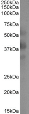
Western Blot using anti-DC-SIGNR (CLEC4M) antibody 16E7 (Ab00130) Jurkat cell extract (35µg protein in RIPA buffer) was resolved on a 10% SDS PAGE gel and blots probed with the chimeric rabbit version of 16E7 (Ab00130-23.0) at 0.1 µg/ml before detection using an anti-rabbit secondary antibody. A primary incubation of 1h was used and protein was detected by chemiluminescence. The expected band size for DC-SIGNR is 45.3kDa, though 9 other isoforms of this protein are known ranging in size from 22.4-44.7kDa (Uniprot ID: Q9H2X3). DC-SIGNR is also glycosylated at several positions. Ab00130-23.0 successfully detected the canonical human DC-SIGNR, as well as multiple other isoforms.