- {{heading}}
- Ab00292-1.1 Anti-IgM [M15/8]
- Human
- Mouse IgG1
- Purified
- In Stock
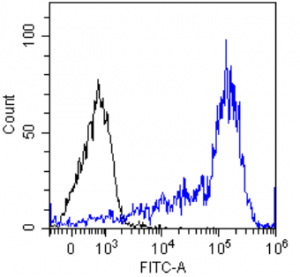
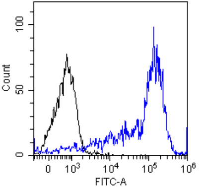
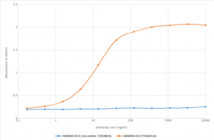
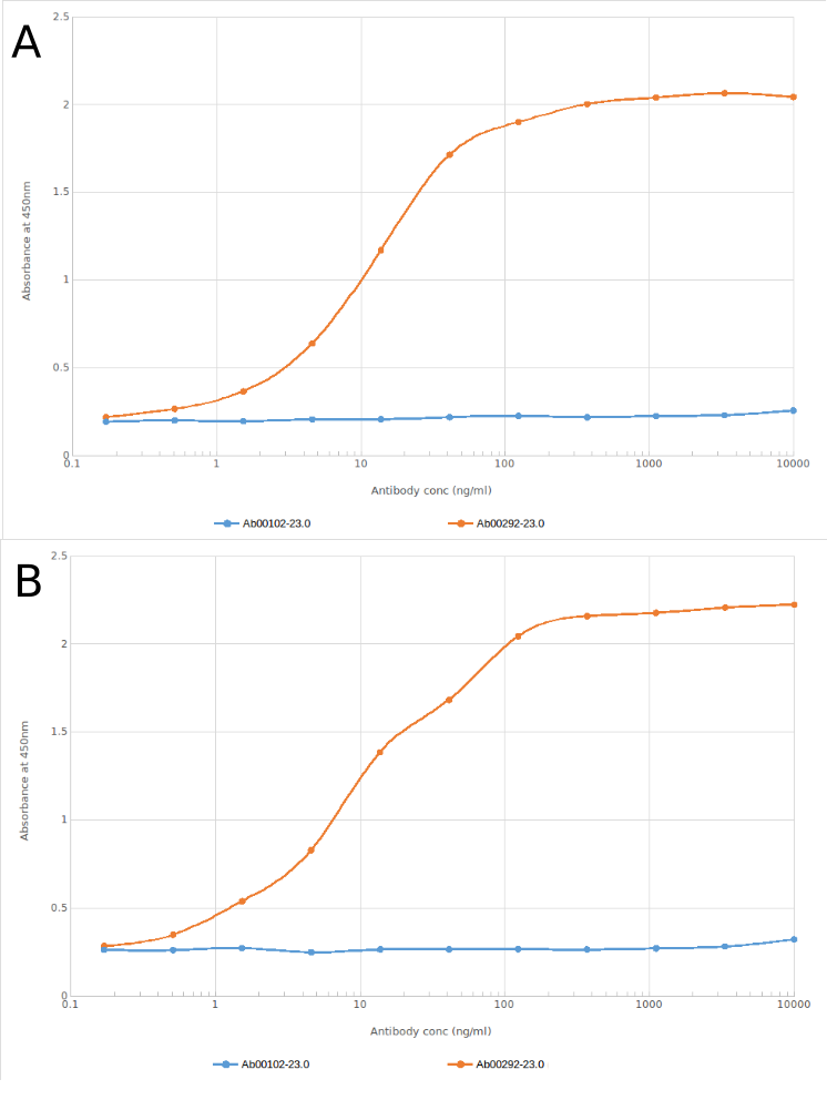
- Ab00292-23.0 Anti-IgM [M15/8]
- Human
- Rabbit IgG
- Purified
- In Stock
- Ab00292-24.1 Anti-IgM [M15/8]
- Human
- Goat IgG
- Purified
- In Stock
Recombinant monoclonal antibody to IgM. Manufactured using AbAb’s Recombinant Platform with variable regions (i.e. specificity) from the hybridoma M15/8.
UniProt Accession Number of Target Protein: P01871
Alternative Name(s) of Target: mu-chain
Immunogen: BALB/c mice immunized with human IgM.
Specificity: This antibody binds to human IgM.
Application Notes: This antibody binds to IgM, which acts in primary immune defines and is involved in early recognition of antigens.
Antibody first published in:
Cragg et al. Analysis of the interaction of monoclonal antibodies with surface IgM on neoplastic B-cells. Br J Cancer. 1999 Feb;79(5-6):850-7 PMID:10070880
Note on publication:
Describes the characterization of the anti-proliferative effect of this antibody on B-cell lymphoma cell lines when applied in a cross-linking F(ab')3 format.
Flow-cytometry using the anti-IgM M15/8 (Ab00292) Daudi cells were stained with unimmunized rabbit IgG antibody (black line) or the rabbit-chimeric version of M15/8 (Ab00292-23.0, blue line) at a concentration of 10 µg/ml for 30 mins at RT. After washing, bound antibody was detected using anti-rabbit IgG JK (FITC-conjugate) antibody (129936) at 2 µg/ml and cells analyzed on a FACSCanto flow-cytometer.
ELISA of anti-IgM antibody on IgM Fc region peptide and a murine IgM antibody. Binding curves of the rabbit chimeric version of the anti-IgM antibody M15/8 (Ab00292-23.0; red line) and isotype control (anti-Fluorescein atnibody; Ab00102-23.0; blue line) to an ELISA plate coated with either IgM Fc region peptide (Pr00108-15.5) at a concentration of 5 µg/ml (A) or murine IgM antibody (B; Anti-CD41 antibody; Ab00222-21.0) again at 5 µg/ml. A 3-fold serial dilution from 10,000 to 0.17 ng/ml was performed using Ab00292-23.0. For signal detection, a 1:4000 dilution of HRP-labelled anti-rabbit IgG1 antibody was used. Both IgM Fc region peptide (monomeric ligand) and murine IgM antibody (multimeric ligand) were recognised by Ab00292-23.0.
Western Blot using anti-IgM antibody M15/8 (Ab00292) Human spleen lysate (35µg protein in RIPA buffer) was resolved on a 10% SDS PAGE gel and blots probed with the chimeric rabbit version of M15/8 (Ab00292-23.0) at 0.3 µg/ml before detection using an anti-rabbit secondary antibody. A primary incubation of 1h was used and protein was detected by chemiluminescence. The expected band size for IgM is 180/900kDa for monomeric/pentameric IgM respectively. Here it is likely that the observed fragment is single glycosylated IgM heavy chain as Ab00292 is targetted to the Fc region of IgM (Cragg et al., PMID: 10070880). Ab00292-23.0 successfully detected IgM in human spleen lysate.
Flow-cytometry using the anti-IgM M15/8 (Ab00292) Daudi cells were stained with unimmunized rabbit IgG antibody (black line) or the rabbit-chimeric version of M15/8 (Ab00292-23.0, blue line) at a concentration of 10 µg/ml for 30 mins at RT. After washing, bound antibody was detected using anti-rabbit IgG JK (FITC-conjugate) antibody (129936) at 2 µg/ml and cells analyzed on a FACSCanto flow-cytometer.
ELISA of anti-IgM antibody on IgM Fc region peptide and a murine IgM antibody. Binding curves of the rabbit chimeric version of the anti-IgM antibody M15/8 (Ab00292-23.0; red line) and isotype control (anti-Fluorescein atnibody; Ab00102-23.0; blue line) to an ELISA plate coated with either IgM Fc region peptide (Pr00108-15.5) at a concentration of 5 µg/ml (A) or murine IgM antibody (B; Anti-CD41 antibody; Ab00222-21.0) again at 5 µg/ml. A 3-fold serial dilution from 10,000 to 0.17 ng/ml was performed using Ab00292-23.0. For signal detection, a 1:4000 dilution of HRP-labelled anti-rabbit IgG1 antibody was used. Both IgM Fc region peptide (monomeric ligand) and murine IgM antibody (multimeric ligand) were recognised by Ab00292-23.0.