2
Products based on this clone:
- {{heading}}
Please Select
- Ab00332-23.0 Anti-PRDX4 [SAIC-40C-8]
- Human
- Rabbit IgG
- Purified
- In Stock
- Ab00332-1.1 Anti-PRDX4 [SAIC-40C-8]
- Human
- Mouse IgG1
- Purified
- Ships in 4-5 weeks
Loading...
 United Kingdom (UK)
Recombinant monoclonal antibody to PRDX4. Manufactured using AbAb’s Recombinant Platform with variable regions (i.e. specificity) from the B-cell cDNA library SAIC-40C-8.
UniProt Accession Number of Target Protein: Q13162
Alternative Name(s) of Target: Peroxiredoxin-4
Immunogen: Peptide "LVQAFQYTDK" derived from the cell redox-regulator Peroxiredoxin-4, conjugated to KLH.
Specificity: Recognises human PRDX4.
Application Notes: Original data characterizing this antibody may be found here .
Antibody first published in:
Schoenherr et al. Anti-peptide monoclonal antibodies generated for immuno-multiple reaction monitoring-mass spectrometry assays have a high probability of supporting Western blot and ELISA. Mol Cell Proteomics PMID:25512614
Note on publication:
Describes generation a panel of recombinant antibodies using synthetic peptides of human proteins as well as validation using ELISA and multiple reaction monitoring-mass spectrometry.
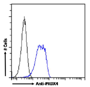
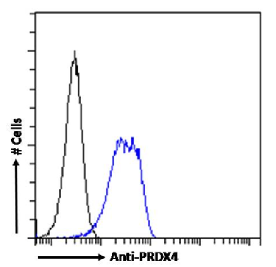
Flow cytometry using the Anti-PRDX4 antibody SAIC-40C-8 (Ab00332). Paraformaldehyde fixed HeLa cells permeabilized with 0.5% Triton were stained with anti-unknown specificity antibody (Ab00178-23.0; isotype control, black line) or the rabbit IgG version of SAIC-40C-8 (Ab00332-23.0, blue line) at a dilution of 1:100 for 1h at RT. After washing, the bound antibody was detected using a goat anti-rabbit IgG AlexaFluor® 488 antibody at a dilution of 1:1000 and cells analyzed using a FACSCanto flow-cytometer.
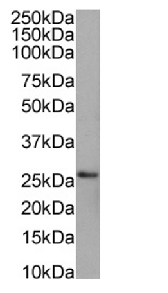
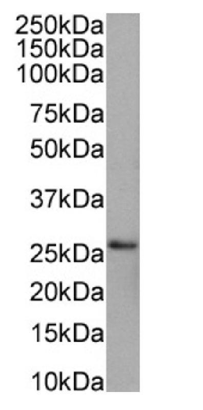
Western Blot using anti-PRDX4 antibody SAIC-40C-8 (Ab00332). HEK293 cell lysates (35µg protein in RIPA buffer) were resolved on a SDS PAGE gel and blots were probed with the chimeric rabbit version of SAIC-40C-8 (Ab00332-23.0) at 0.001 µg/ml before detection using an anti-rabbit secondary antibody. A primary incubation of 1h was used and protein was detected by chemiluminescence.
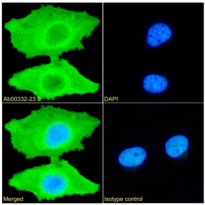
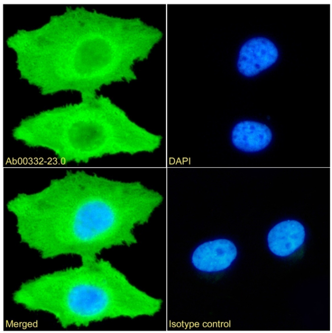
Immunofluorescence staining of HeLa cells with anti-PRDX4 (Ab00332) SAIC-40C-8 Immunofluorescence analysis of paraformaldehyde fixed HeLa cells permeabilized with 0.15% Triton stained with the chimeric rabbit IgG version of SAIC-40C-8 (Ab00332-23.0) (1:100 dilution) for 1h followed by Alexa Fluor® 488 secondary antibody (1:1000 dilution), showing cytoplasmic staining. The nuclear stain is DAPI (blue). Panels show from left-right, top-bottom Ab00332-23.0, DAPI, merged channels and an isotype control. The isotype control was an unknown specificity antibody (Ab00178-23.0) followed by staining with Alexa Fluor® 488 secondary antibody.