- {{heading}}
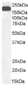
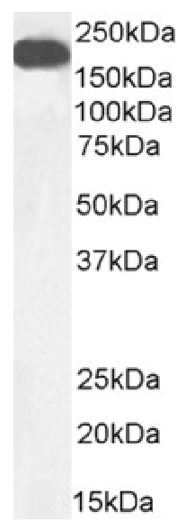
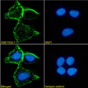
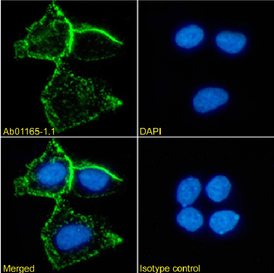
- Ab01165-1.1 Anti-carcinoembryonic antigen [A5B7]
- Human
- Mouse IgG1
- Purified
- In Stock
- Ab01165-23.0 Anti-carcinoembryonic antigen [A5B7]
- Human
- Rabbit IgG
- Purified
- In Stock
Recombinant monoclonal antibody to carcinoembryonic antigen. Manufactured using AbAb’s Recombinant Platform with variable regions (i.e. specificity) from the hybridoma A5B7.
UniProt Accession Number of Target Protein: Q13982
Alternative Name(s) of Target: CEA; CD66; Q7KZ28; Q13985; Q14081
Immunogen: This antibody was raised by immunising mice with both carcinoembryonic antigen (CEA) purified from the metastases of colonic tumours, and heat-treated CEA.
Specificity: This antibody is specific for human CEA.
Application Notes: In IHC analysis, this CEA-specific antibody stains sections of colon tumour, with high levels of cell surface and cytoplasmic staining of malignant glands, and a more intense reaction in necrotic debris (Harwood et al, 1986). It does not cross-react with PMNs, erythrocytes or liver tissue. In an mouse model transplanted with the human colonic tumour MaWi, the weight of the xenograft has a profound effect on the uptake of a radiolabelled version of antibody (Pedley et al, 1987). In a xenograft tumour mouse model based on the adenocarcinoma cell line LS174T, both this antibody and its FAb have a significant effect on tumour growth when administered as radioimmunotherapeutic agents (Pedley et al, 1993). This antibody has also been shown to function as a radioimmunotherapy agent in a Phase I trial in gastrointestinal adenocarcinomas (Meyer et al, 2009), and has been shown in early clinical trials to target an enzyme capable of cleaving an inactive prodrug to release an active, cytotoxic drug to colorectal cancers (Bagshawe et al, 1991).
Antibody first published in:
Harwood et al. Mapping epitope characteristics on carcinoembryonic antigen. Br J Cancer. 1986 Jul;54(1):75-82 PMID:2425837
Note on publication:
Describes the original generation of this antibody, and its characterisation in IHC analysis.
Western Blot using anti-carcinoembryonic antigen antibody A5B7 (Ab01165). human colorectal carcinoma tissue lysate (35µg protein in RIPA buffer) was resolved on an SDS PAGE gel and blots probed with the chimeric mouse IgG version of A5B7 (Ab01165-1.1) at 0.01 µg/ml before detection using an anti-mouse secondary antibody. A primary incubation of 1h was used and protein was detected by chemiluminescence.
Immunofluorescence staining of MCF7 cells using anti-carcinoembryonic antigen (Ab01165) A5B7 Immunofluorescence analysis of paraformaldehyde fixed MCF7 cells stained with the chimeric mouse IgG version of A5B7 (Ab01165-1.1) at 10 µg/ml followed by Alexa Fluor® 488 secondary antibody (2 µg/ml), showing membrane staining. The nuclear stain is DAPI (blue). Panels show from left-right, top-bottom Ab01215-1.1, DAPI, merged channels and an isotype control. The isotype control was stained with anti-unknown antibody (Ab00178-1.1) followed by Alexa Fluor® 488 secondary antibody.
Western Blot using anti-carcinoembryonic antigen antibody A5B7 (Ab01165). human colorectal carcinoma tissue lysate (35µg protein in RIPA buffer) was resolved on an SDS PAGE gel and blots probed with the chimeric mouse IgG version of A5B7 (Ab01165-1.1) at 0.01 µg/ml before detection using an anti-mouse secondary antibody. A primary incubation of 1h was used and protein was detected by chemiluminescence.
Flow cytometry using the Anti-carcinoembryonic antigen antibody A5B7 (Ab01165). Paraformaldehyde fixed MCF7 cells were stained with anti-unknown specificity antibody (Ab00178-1.1; isotype control, black line) or the mouse IgG1 version of A5B7 (Ab01165-1.1, blue line) at a dilution of 1:100 for 1h at RT. After washing, the bound antibody was detected using a goat anti-mouse IgG AlexaFluor® 488 antibody at a dilution of 1:1000 and cells analyzed using a FACSCanto flow-cytometer.