- {{heading}}
- Ab01444-34.11 Anti-IgG1 Fc [TP894]
- Mouse
- Camelid VHH
- His-Tagged
- Purified
- In Stock
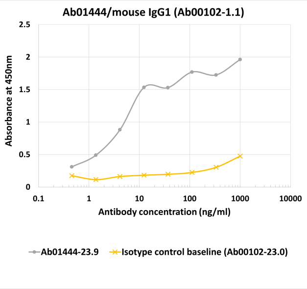
- Ab01444-23.9 Anti-IgG1 Fc [TP894]
- Mouse
- Rabbit IgG-Fc fusion
- His-Tagged
- Purified
- In Stock
Recombinant monoclonal antibody to IgG1 Fc. Manufactured using AbAb’s Recombinant Platform with variable regions (i.e. specificity) from the phage display antibody TP894.
Alternative Name(s) of Target: immunoglobulin G1 fragment crystallizable region; immunoglobulin G1 Fc region; immunoglobulin G1
Immunogen: This antibody was raised by immunising alpacas with 1.0 mg polyclonal mouse IgG. Subsequently, the generation of nanobody immune libraries and the selection of antigen-specific nanobodies by phage display from these libraries were performed.
Specificity: This antibody recognises Fc fragment of the IgG1.
Application Notes: Anti-IgG nanobodies often perform more efficiently than polyclonal secondary antibodies in various experimental assays. Therefore, they are recommended for various experimental procedures. Anti-IgG nanobodies were shown to be more sensitive during Western blots when conjugated with different molecules (HRP, fluorophores etc.) (Pleiner et al., 2018). Furthermore, combination of two distinct nanobodies for immunofluorescence labelling provided exceptionally clean and strong signal, unprecedented among monovalent secondary antibodies (Pleiner et al., 2018). Additionally, using such an antibody allows skipping two-step incubation procedures during immunostaing as the nanobody can be first incubated with a primary antibody and this conjugate might be added to the cells in a one-step procedure (Pleiner et al., 2018). Finally, such nanobodies have been recommended as very effective for colocalisation studies when more than one fluorophore is needed (Pleiner et al., 2018).
Antibody first published in:
Pleiner et al. A toolbox of anti–mouse and anti–rabbit IgG secondary nanobodies. J Cell Biol. 2018 Mar 5;217(3):1143-1154. doi: 10.1083/jcb.201709115. Epub 2017 Dec 20. PMID:29263082
Note on publication:
The article describes the generation and characterisation of this nanobody.
ELISA targeting mouse IgG1 using anti-mouse IgG1 Fc antibody TP894 (Ab01444). Binding of the chimeric rabbit IgG-Fc fusion (his-tagged) version of the anti-mouse IgG1 Fc antibody TP894 (Ab01444-23.9; gray line), and the appropriate isotype control (anti-fluorescein 4-4-20 (enhanced); Ab00102-23.0, yellow line), to mouse IgG1 was measured in an ELISA from 1000 to 0.46 ng/ml concentration in 3-fold serial dilution steps. The reaction plate was coated with the chimeric mouse IgG1 version of anti-fluorescein 4-4-20 (enhanced) (Ab00102-1.1) at a 2.5 µg/ml concentration. A 1:4000 dilution of an HRP-conjugated anti-rabbit IgG antibody was used for signal detection.