2
Products based on this clone:
- {{heading}}
Please Select
- Ab00215-1.1 Anti-MUC1 [SM3]
- Human
- Mouse IgG1
- Purified
- In Stock
- Ab00215-23.0 Anti-MUC1 [SM3]
- Human
- Rabbit IgG
- Purified
- In Stock
Loading...
 United Kingdom (UK)
Recombinant monoclonal antibody to MUC1. Manufactured using AbAb’s Recombinant Platform with variable regions (i.e. specificity) from the hybridoma SM3.
UniProt Accession Number of Target Protein: P15941
Alternative Name(s) of Target: Mucin
Immunogen: Hydrogen fluoride deglycosylated milk mucin.
Specificity: Recognises the under-glycosylated form of human MUC1, a marker of tumours.
Application Notes: This antibody binds to mucin, a heavily glycosylated protein produced by epithelial cells that forms gels.
Antibody first published in: Burchell J, Gendler S, Taylor-Papadimitriou J, Girling A, Lewis A, Millis R, Lamport D. Development and characterization of breast cancer reactive monoclonal antibodies directed to the core protein of the human milk mucin. Cancer Res. 1987 Oct 15;47(20):5476-82. PMID:2443241
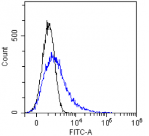
Flow-cytometry using the anti-MUC1 SM3 (Ab00215) MCF-7 cells were stained with unimmunized rabbit IgG antibody (black line) or the rabbit-chimeric version of SM3 (Ab00215-23.0, blue line) at a concentration of 10 µg/ml for 30 mins at RT. After washing, bound antibody was detected using anti-rabbit IgG JK (FITC-conjugate) antibody (129936) at 2 µg/ml and cells analyzed on a FACSCanto flow-cytometer.
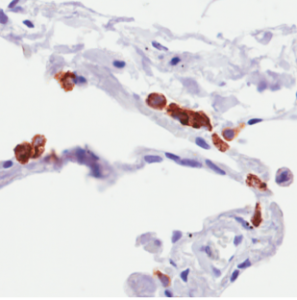
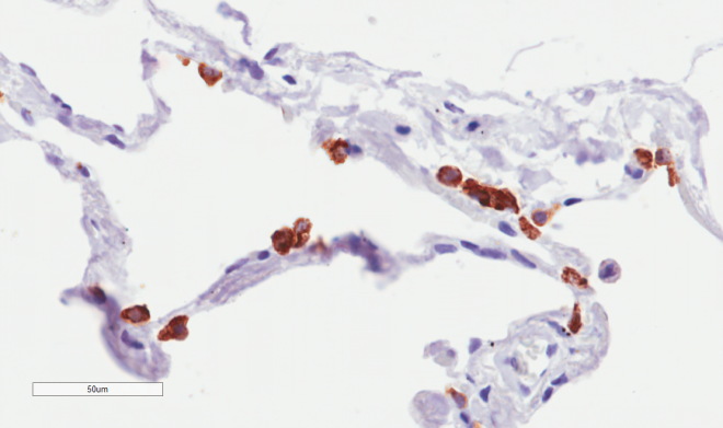
Immunohistochemical staining of human lung tissue using anti-MUC1 antibody (Ab00215) SM3 Anti-MUC1 (Mucin-1) staining of paraffin embedded human lung tissue using the rabbit-chimeric version of SM3 (Ab00215-23.0). Antigen retreival was acheived by microwaving in citrate buffer (pH6), followed by blocking with protein block serum-free buffer (Dako, cat. #X0909). Primary antibody incubation with Ab00215-23.0 was carried out at 4 µg/ml for 30 minutes. Samples were then incubated with an anti-rabbit IgG HRP secondary antibody (Dako cat#K4009) for 20 mins followed by DAB (3,3′-diaminobenzidine), and counter-staining with haemotoxylin. Staining of type II pneumocytes may be observed. Recommended concentration, 2-4 µg/ml.
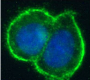
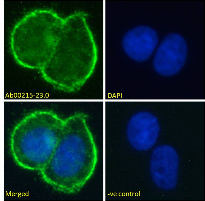
Immunofluorescence staining of fixed MCF-7 cells with anti-MUC1 antibody SM3 (Ab00215) Immunofluorescence analysis of unpermeabilisd paraformaldehyde fixed MCF-7 cells on Shi-fix™ coverslips stained with the chimeric rabbit version of SM3 (Ab00215-23.0) at 10 µg/ml for 1h followed by Alexa Fluor® 488 secondary antibody (1 µg/ml), showing clear membrane staining. The nuclear stain is DAPI (blue). Panels show from left-right, top-bottom; Ab00215-23.0, DAPI, merged channels and an isotype control. The isotype control was stained with unimmunised rabbit IgG followed by Alexa Fluor® 488 secondary antibody.
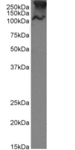
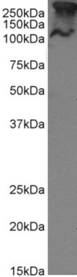
Western Blot using anti-MUC1 antibody SM3 (Ab00215) MCF-7 cell lysate (35µg protein in RIPA buffer) were resolved on a 10% SDS PAGE gel and blots probed with the chimeric rabbit version of SM3 (Ab00215-23.0) at 1 µg/ml before detection using an anti-rabbit secondary antibody. A primary incubation of 1h was used and protein was detected by chemiluminescence. The predicted band size for unmodified MUC1 is 122.1kDa, though in breast cancer cell lines like MCF-7 MUC1 can be up to 90% glycosylated (c.f. Mueller et al. PMID: 10373415; T47D cells) and expected band sizes are ~250-300kDa. Thus the two bands likely represent processed (>250kDa) and unprocessed (~121kDa) populations of the protein. Ab00215-23.0 successfully detected human MUC1 in MCF-7 breast cancer cells.
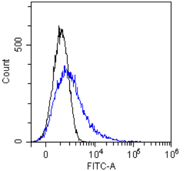
Flow-cytometry using the anti-MUC1 SM3 (Ab00215) MCF-7 cells were stained with unimmunized rabbit IgG antibody (black line) or the rabbit-chimeric version of SM3 (Ab00215-23.0, blue line) at a concentration of 10 µg/ml for 30 mins at RT. After washing, bound antibody was detected using anti-rabbit IgG JK (FITC-conjugate) antibody (129936) at 2 µg/ml and cells analyzed on a FACSCanto flow-cytometer.