- {{heading}}
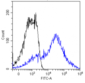
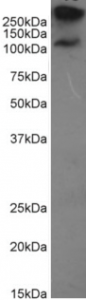
- Ab00712-1.1 Anti-MUC1 [HMFG2]
- Human
- Mouse IgG1
- Purified
- In Stock
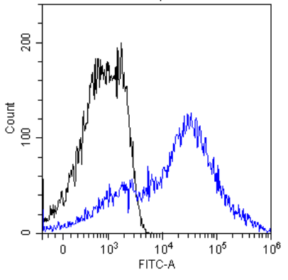
- Ab00712-1.4 Anti-MUC1 [HMFG2]
- Human
- Mouse IgG1
- Fc Silent™
- Purified
- Ships in 4-5 weeks
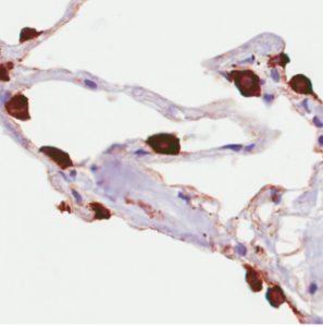
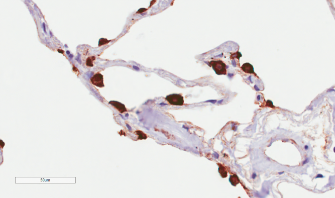
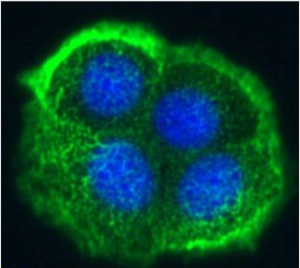
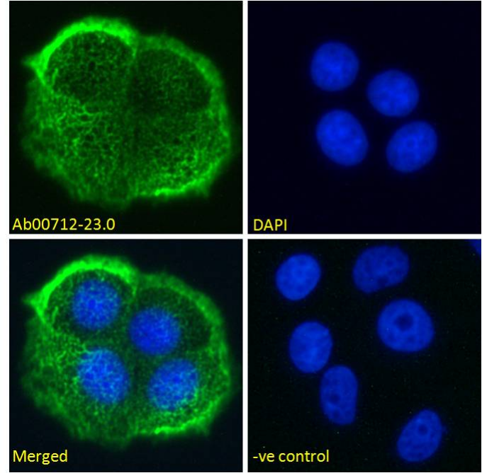
- Ab00712-23.0 Anti-MUC1 [HMFG2]
- Human
- Rabbit IgG
- Purified
- In Stock
Recombinant monoclonal antibody to MUC1. Manufactured using AbAb’s Recombinant Platform with variable regions (i.e. specificity) from the hybridoma HMFG2.
UniProt Accession Number of Target Protein: P15941
Alternative Name(s) of Target: Muc-1
Immunogen: High-MW glycoprotein of the human milk fat globule.
Specificity: This antibody recognises MUC1, a tumor associated antigen which is expressed in >90% ovarian carcinomas. This antigen is a high molecular weight (M, 80,000-200,000) glycoprotein.
Application Notes: This antibody binds to MUC1, an antigen widely expressed on breast cancer cells. This antibody antibodies react strongly with malignant epithelial cells but not with normal mesothelial or endothelial cells.
Antibody first published in:
Taylor-Papadimitriou Monoclonal antibodies to epithelium-specific components of the human milk fat globule membrane: production and reaction with cells in culture. Int J Cancer. 1981 Jul 15;28(1):17-21. PMID:7309278
Note on publication:
Describes the generation and initial characterisation of the specificity of this antibody to breast cancer cells.
Flow-cytometry using the anti-MUC1 antibody HMFG2 (Ab00712) MCF-7 cells were stained with unimmunized rabbit IgG antibody (black line) or the rabbit-chimeric version of HMFG2 (Ab00712-23.0, blue line) at a concentration of 10 µg/ml for 30 mins at RT. After washing, bound antibody was detected using an anti-rabbit IgG JK (FITC-conjugate) antibody (129936) at 2 µg/ml and cells analyzed on a FACSCanto flow-cytometer.
Immunohistochemical staining of human lung tissue using anti-MUC1 antibody (Ab00712) HMFG2 Anti-MUC1 (Mucin-1) staining of paraffin embedded human lung tissue using the rabbit-chimeric version of HMFG2 (Ab00712-23.0). Antigen retreival was acheived by microwaving in citrate buffer (pH6), followed by blocking with protein block serum-free buffer (Dako, cat. #X0909). Primary antibody incubation with Ab00712-23.0 was carried out at 4 µg/ml for 30 minutes. Samples were then incubated with an anti-rabbit IgG HRP secondary antibody (Dako cat#K4009) for 20 mins followed by DAB (3,3′-diaminobenzidine), and counter-staining with haemotoxylin. Strong staining of type II pneumocytes may be observed. Recommended concentration, 1-2 µg/ml.
Immunofluorescence staining of fixed MCF-7 cells with anti-MUC1 antibody HMFG2 (Ab00712) Immunofluorescence analysis of unpermeabilisd paraformaldehyde fixed MCF-7 cells on Shi-fix™ coverslips stained with the chimeric rabbit version of HMFG2 (Ab00712-23.0) at 10 µg/ml for 1h followed by Alexa Fluor® 488 secondary antibody (1 µg/ml), showing membrane staining. The nuclear stain is DAPI (blue). Panels show from left-right, top-bottom Ab00712-23.0, DAPI, merged channels and an isotype control. The isotype control was stained with an anti-Fluorescein antibody (Ab102-23.0) followed by Alexa Fluor® 488 secondary antibody.
Western Blot using anti-MUC1 antibody HMFG2 (Ab00712) MCF-7 cell lysate (35µg protein in RIPA buffer) were resolved on a 10% SDS PAGE gel and blots probed with the chimeric rabbit version of HMFG2 (Ab00712-23.0) at 0.1 µg/ml before detection using an anti-rabbit secondary antibody. A primary incubation of 1h was used and protein was detected by chemiluminescence. The predicted band size for unmodified MUC1 is 122.1kDa, though in breast cancer cell lines like MCF-7 MUC1 can be up to 90% glycosylated (c.f. Mueller et al. PMID: 10373415; T47D cells) and expected band sizes are ~250-300kDa. Thus the two bands likely represent processed (>250kDa) and unprocessed (~121kDa) populations of the protein. Ab00712-23.0 successfully detected human MUC1 in MCF-7 breast cancer cells.
Flow-cytometry using the anti-MUC1 antibody HMFG2 (Ab00712) MCF-7 cells were stained with unimmunized rabbit IgG antibody (black line) or the rabbit-chimeric version of HMFG2 (Ab00712-23.0, blue line) at a concentration of 10 µg/ml for 30 mins at RT. After washing, bound antibody was detected using an anti-rabbit IgG JK (FITC-conjugate) antibody (129936) at 2 µg/ml and cells analyzed on a FACSCanto flow-cytometer.