- {{heading}}
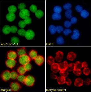
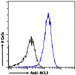
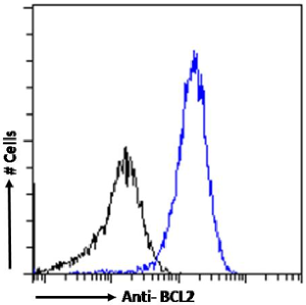
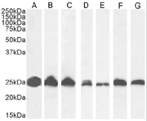
- Ab01321-1.1 Anti-BCL2 [bcl-2/100]
- Human
- Mouse IgG1
- Purified
- In Stock
- Ab01321-23.0 Anti-BCL2 [bcl-2/100]
- Human
- Rabbit IgG
- Purified
- In Stock
- Ab01321-10.0 Anti-BCL2 [bcl-2/100]
- Human
- Human IgG1
- Purified
- Ships in 4-5 weeks
Recombinant monoclonal antibody to BCL2. Manufactured using AbAb’s Recombinant Platform with variable regions (i.e. specificity) from the hybridoma bcl-2/100.
UniProt Accession Number of Target Protein: P10415
Alternative Name(s) of Target: Apoptosis regulator Bcl-2; AW986256; B-cell CLL/lymphoma 2; Bcl-2; C430015F12Rik; D630044D05Rik; D830018M01Rik; protein phosphatase 1; regulatory subunit 50; bcl-2-100; bcl2/100
Immunogen: This antibody was raised against a synthetic peptide of amino acids 41-54 of human bcl-2 oncoprotein.
Specificity: This antibody recognises human BLC-2 which is an antiapoptotic factor in various cell types. Its disregulation was implicated in different conditions, such as cancers and autoimmune disorders.
Application Notes: Bcl-2 has been implicated in various cancers, such as lymphoma, melanoma or colorectal cancer. Therefore, multiple immunohistochemical stainings against bcl-2 protein were performed and this antibody was found to be useful in characterisation of molecular features of certain cancers. ICH: One group used bcl-2/100 antibody to stain paraffin-embedded colorectal cancer samples and strong signal, mainly from the colonic cells, was observed in the tissue (Nicholson et al., 2014). Another group noted bcl-2 immunohistochemical staining in both morular and nonmorular papillary thyroid carcinoma samples while trying to identify cliinicopathological features of this carcinoma (Jung et al., 2009). Finally, yet another found that neoplastic cells of the primary sclerosing epithelioid fibrosarcoma (SEF) of the kidney are characterised by a strong anti-bcl2 immunohistochemical staining with this antibody (Ertoy Baydar et al., 2015). WB: Additionally, multiple groups were successfull while using bcl-2/100 antibody to detect bcl-2 via Western blot; for example in HEC cells while investigating the influence of simvastatin combined with kallistatin on paediatric burn patients with sepsis (Dai and Zhao, 2018).
Antibody first published in:
Pezzella et al. Expression of the bcl-2 oncogene protein is not specific for the 14;18 chromosomal translocation. Am J Pathol. 1990 Aug;137(2):225-32. PMID:2201196
Note on publication:
This article describes generation and characterisation of the bcl-2/100 antibody.
Immunofluorescence staining of Jurkat cells using anti-BCL2 antibody (Ab01321) bcl-2/100. Immunofluorescence analysis of paraformaldehyde fixed Jurkat cells permeabilized with 0.15% Triton and stained with the chimeric mouse IgG version of bcl-2/100 (Ab01321-1.1) at 10 µg/ml followed by Alexa Fluor® 488 secondary antibody (2 µg/ml), showing nuclear staining. Actin filaments were stained with phalloidin (red) and the nuclear stain is DAPI (blue). Panels show from left-right, top-bottom Ab01321-1.1, DAPI, merged channels and an isotype control. The isotype control was stained with anti-unknown antibody (Ab00178-1.1) followed by Alexa Fluor® 488
Flow-cytometry using the anti-BCL2 antibody bcl-2/100 (Ab01321). Jurkat cells were fixed using paraformaldehyde, permeabilised using 0.5% Triton and stained with anti-unknown IgG antibody (Ab00178-1.1; isotype control, black line) or the mouse IgG-chimeric version of bcl-2/100 (Ab01321-1.1, blue line) at a dilution of 1:100 for 1h at RT. After washing, bound antibody was detected using a goat anti-mouse IgG AlexaFluor® 488 antibody at a dilution of 1:1000 and cells analyzed using a FACSCanto flow-cytometer.
Western Blot using anti-BCL2 antibody bcl-2/100 (Ab01321). Jurkat(A) (0.01 µg/ml), U937(B) (0.01 µg/ml), K562(C) (0.03µg/ml), HeLa(D) cells (0.1 µg/ml), human spleen(E) (0.3 µg/ml), human thymus(F) (0.03µg/ml) and human tonsil(G) (0.03 µg/ml) tissue lysate (35µg protein in RIPA buffer) was resolved on an SDS PAGE gel and blots probed with the chimeric mouse IgG version of bcl-2/100 (Ab01321-1.1) before detection using an anti-mouse secondary antibody. A primary incubation of 1h was used and protein was detected by chemiluminescence.
Flow cytometry using the Anti-BCL2 antibody bcl-2/100 (Ab01321). Paraformaldehyde fixed Jurkat cells permeabilized with 0.5% Triton were stained with anti-unknown specificity antibody (Ab00178-1.1; isotype control, black line) or the mouse IgG1 version of bcl-2/100 (Ab01321-1.1, blue line) at a dilution of 1:100 for 1h at RT. After washing, the bound antibody was detected using a goat anti-mouse IgG AlexaFluor® 488 antibody at a dilution of 1:1000 and cells analyzed using a FACSCanto flow-cytometer.