- {{heading}}
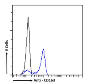
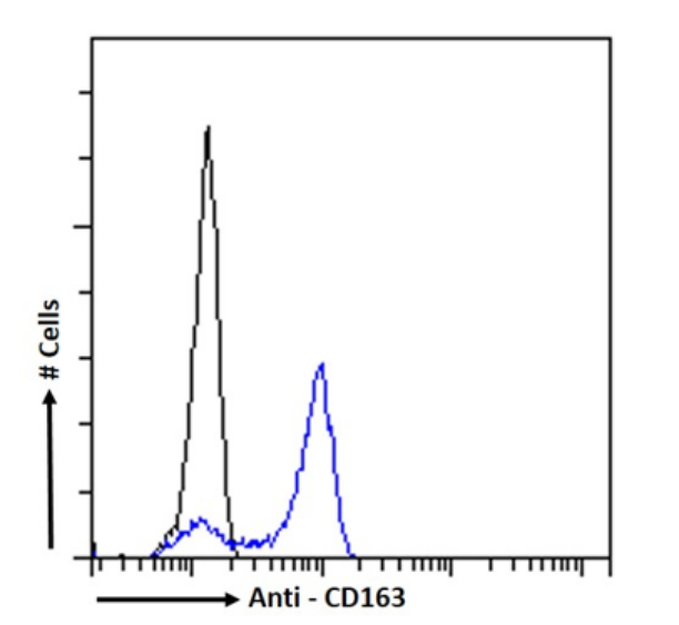
- Ab02085-1.1 Anti-CD163 [2A10 (2A10/11)]
- Pig (Sus scrofa)
- Mouse IgG1
- Purified
- In Stock
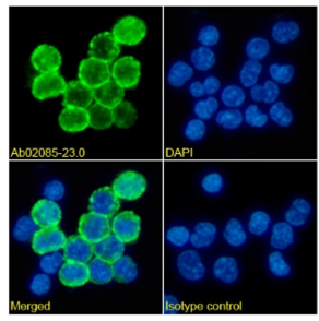
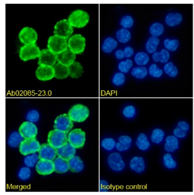
- Ab02085-23.0 Anti-CD163 [2A10 (2A10/11)]
- Pig (Sus scrofa)
- Rabbit IgG
- Purified
- In Stock
Recombinant monoclonal antibody to CD163. Manufactured using AbAb’s Recombinant Platform with variable regions (i.e. specificity) from the hybridoma 2A10 (2A10/11).
UniProt Accession Number of Target Protein: Q2VL90
Alternative Name(s) of Target: M130; sCD163; Scavenger receptor cysteine-rich type 1 protein M130
Immunogen: The original antibody was generated by immunizing BALB/c mice with porcine alveolar macrophages.
Specificity: This antibody recognizes and binds porcine CD163.
Application Notes: This antibody is recommended to be used as a tool in the study of porcine macrophage maturation and differentiation and for determining their heterogeneity both in normal and pathological conditions. This antibody can recognize porcine CD163 in a western blot assay and an immunoprecipitation assay (PMID: 9151393). 2A10 successfully binds PAM cells in a flow cytometric analysis (PMID:23785195). Porcine CD163 has also been analyzed as potential antigen targeting receptors using mouse IgG as surrogate antigens. MAbs to CD163 induced in vitro PBMC proliferation at concentrations 50-80 fold lower than the control mAb when using, as responder cells, cells from pigs immunized with mouse serum IgGs (PMID:21557980). It is also reported that porcine mAb 2A10 antigen is homologous to human CD163 and related to macrophage differentiation (PMID: 10227997). This antibody was also used to study the role of CD163 in the process of infection of porcine monocytes/macrophages by African swine fever virus infection (PMID: 14648288). This mAb was used in a sandwich enzyme-linked immunoassay (ELISA) to measure levels of sCD163 in porcine sera and biological fluids (Pérez et al, 2008).
Antibody first published in:
Bullido et al. Monoclonal antibodies specific for porcine monocytes/macrophages: macrophage heterogeneity in the pig evidenced by the expression of surface antigens. Tissue Antigens (1997); 49(4):403-13. PMID:9151393
Note on publication:
Describes the distribution of surface antigens on porcine monocytes and macrophages using monoclonal antibodies.
Immunofluorescence staining of pig peripheral blood monocytes with anti-CD163 (Ab02085) 2A10 (2A10/11) Immunofluorescence analysis of paraformaldehyde fixed pig peripheral blood monocytes on Shi-fix™ coverslips stained with the chimeric rabbit IgG version of 2A10 (2A10/11) (Ab02085-23.0) at 10 µg/ml for 1h followed by Alexa Fluor® 488 secondary antibody (2 µg/ml), showing membrane staining. The nuclear stain is DAPI (blue). Panels show from left-right, top-bottom Ab02085-23.0, DAPI, merged channels and an isotype control. The isotype control was an unknown specificity antibody (Ab00178-23.0) followed by staining with Alexa Fluor® 488 secondary antibody.
Flow cytometry using the Anti-CD163 antibody 2A10 (2A10/11) (Ab02085). Paraformaldehyde fixed pig peripheral blood monocytes permeabilized with 0.5% Triton were stained with anti-unknown specificity antibody (Ab00178-23.0; isotype control, black line) or the rabbit IgG version of 2A10 (2A10/11) (Ab02085-23.0, blue line) at a dilution of 1:100 for 1h at RT. After washing, the bound antibody was detected using a goat anti-rabbit IgG AlexaFluor® 488 antibody at a dilution of 1:1000 and cells analyzed using a FACSCanto flow-cytometer.
Immunofluorescence staining of pig peripheral blood monocytes with anti-CD163 (Ab02085) 2A10 (2A10/11) Immunofluorescence analysis of paraformaldehyde fixed pig peripheral blood monocytes on Shi-fix™ coverslips stained with the chimeric rabbit IgG version of 2A10 (2A10/11) (Ab02085-23.0) at 10 µg/ml for 1h followed by Alexa Fluor® 488 secondary antibody (2 µg/ml), showing membrane staining. The nuclear stain is DAPI (blue). Panels show from left-right, top-bottom Ab02085-23.0, DAPI, merged channels and an isotype control. The isotype control was an unknown specificity antibody (Ab00178-23.0) followed by staining with Alexa Fluor® 488 secondary antibody.