2
Products based on this clone:
- {{heading}}
Please Select
- Ab01427-1.1 Anti-CD171 [L1-14.10]
- Human
- Mouse IgG1
- Purified
- In Stock
- Ab01427-23.0 Anti-CD171 [L1-14.10]
- Human
- Rabbit IgG
- Purified
- In Stock
Loading...
 United Kingdom (UK)
Recombinant monoclonal antibody to CD171. Manufactured using AbAb’s Recombinant Platform with variable regions (i.e. specificity) from the hybridoma L1-14.10.
UniProt Accession Number of Target Protein: P32004
Alternative Name(s) of Target: L1CAM; 14.10; L1; Neural cell adhesion molecule L1; N-CAM-L1; NCAM-L1
Immunogen: This antibody was raised by immunising mice with recombinant L1-Fc fusion protein, consisting of the ectodomain of human L1.
Specificity: This antibody is specific for the ectodomain (amino acids M1 to E1123) of human L1, and recognises an epitope in the third Ig domain. L1 is over-expressed in a range of tumours, including neuroblastomas, renal carcinomas, ovarian and endometrial carcinomas, and melanomas. This antibody does not cross-react with human CHL1.
Application Notes: The specificity of this antibody has been confirmed in flow cytometric (Schafer et al, 2012; Huszar et al, 2006) and Western blot analysis (Huszar et al, 2006; Wolterink et al, 2010). This antibody has been shown, when incubated with SKOV3ip human ovarian carcinoma cells, to efficiently and sustainedly block extracellular signal-regulated kinase (Erk) phosphorylation (Gast et al, 2008). This antibody has been used in immunohistochemical staining for L1 expression, using paraffin-embedded human ovarian carcinoma tissue sections (Wolterink et al, 2010; Knogler et al, 2007) and a panel of human tumour tissue sections, including benign and malignant tumours of the female genital tract (Huszar et al, 2006), and renal cell carcinoma, schwannoma, gastric conventional GIST, pancreatic neuroendocrine tumor and paraganglioma (Inaguma et al, 2016). This antibody, when administered to SCID mice in combination with cytostatic drugs, efficiently reduces the growth of subcutaneously grown Colo357 or SKOV3ip tumors (Schafer et al, 2012).
Antibody first published in:
Huszar et al. Expression profile analysis in multiple human tumors identifies L1 (CD171) as a molecular marker for differential diagnosis and targeted therapy. Hum Pathol. 2006 Aug;37(8):1000-8. Epub 2006 Jun 21. PMID:16867862
Note on publication:
Describes the original generation of this antibody, and its use in immunohistochemical analysis.
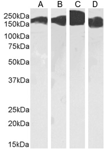
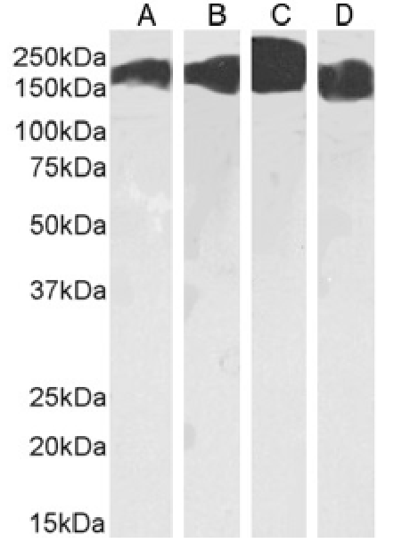
Western Blot using anti-CD171 antibody L1-14.10 (Ab01427). HeLa(A) (0.03 µg/ml), Kelly(B) cells (0.01 µg/ml), human brain cerebellum(C) (0.003 µg/ml) and cerebral cortex(D) (0.01 µg/ml) tissue lysate (35µg protein in RIPA buffer) was resolved on an SDS PAGE gel and blots probed with the chimeric mouse IgG version of L1-14.10 (Ab01427-1.1) before detection using an anti-mouse secondary antibody. A primary incubation of 1h was used and protein was detected by chemiluminescence.
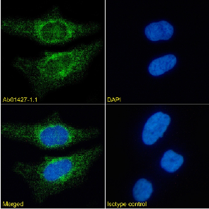
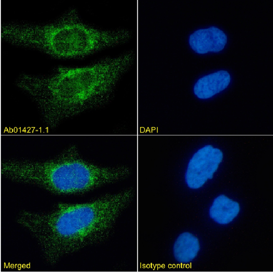
Immunofluorescence staining of HeLa cells using anti-CD171 (Ab01427) L1-14.10 Immunofluorescence analysis of paraformaldehyde fixed HeLa cells stained with the chimeric mouse IgG version of L1-14.10 (Ab01427-1.1) at 10 µg/ml followed by Alexa Fluor® 488 secondary antibody (2 µg/ml), showing cytoplasmic staining. The nuclear stain is DAPI (blue). Panels show from left-right, top-bottom Ab01215-1.1, DAPI, merged channels and an isotype control. The isotype control was stained with anti-unknown antibody (Ab00178-1.1) followed by Alexa Fluor® 488 secondary antibody.
Western Blot using anti-CD171 antibody L1-14.10 (Ab01427). HeLa(A) (0.03 µg/ml), Kelly(B) cells (0.01 µg/ml), human brain cerebellum(C) (0.003 µg/ml) and cerebral cortex(D) (0.01 µg/ml) tissue lysate (35µg protein in RIPA buffer) was resolved on an SDS PAGE gel and blots probed with the chimeric mouse IgG version of L1-14.10 (Ab01427-1.1) before detection using an anti-mouse secondary antibody. A primary incubation of 1h was used and protein was detected by chemiluminescence.
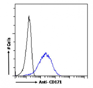
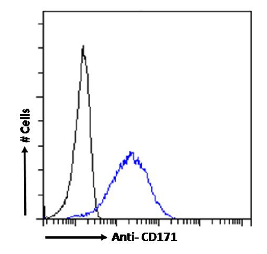
Flow cytometry using the Anti-CD171 antibody L1-14.10 (Ab01427). Paraformaldehyde fixed HeLa cells were stained with anti-unknown specificity antibody (Ab00178-1.1; isotype control, black line) or the mouse IgG1 version of L1-14.10 (Ab01427-1.1, blue line) at a dilution of 1:100 for 1h at RT. After washing, the bound antibody was detected using a goat anti-mouse IgG AlexaFluor® 488 antibody at a dilution of 1:1000 and cells analyzed using a FACSCanto flow-cytometer.