2
Products based on this clone:
- {{heading}}
Please Select
- Ab00289-1.1 Anti-CD38 [AT13/5]
- Human
- Mouse IgG1
- Purified
- In Stock
- Ab00289-23.0 Anti-CD38 [AT13/5]
- Human
- Rabbit IgG
- Purified
- In Stock
Loading...
 United Kingdom (UK)
Recombinant monoclonal antibody to CD38. Manufactured using AbAb’s Recombinant Platform with variable regions (i.e. specificity) from the hybridoma AT13/5.
UniProt Accession Number of Target Protein: P28907
Alternative Name(s) of Target: ADP-ribosyl cyclase 1; cyclic ADP-ribose hydrolase; EC 3.2.2.5; Cyclic ADP-ribose hydrolase 1; cADPr hydrolase 1; T10; CD38; p45; NAD(+) nucleosidase
Immunogen: Namalwa cells emulsified in CFA.
Specificity: Binds specifically to human CD38, an approximately 45 kDa type II transmembrane protein, expressed on essentially all pre-B lymphocytes, plasma cells, and thymocytes. Also present on activated T lymphocytes, natural killer (NK) lymphocytes, myeloblasts, and erythroblasts. Bimodally expressed during B cell development, modulating from high in immature cells to low in intermediate ones and back to high on mature B cells. This antibody competes with clone HB7 (Ab00128) (Ellis 1995) which has been shon to bind to an epitope between amino acids 273-285).
Application Notes: The antibody binds to CD38, a glycoportein on the surface of various immune cells that catalyzes the hydrolysis of cyclic ADP-ribose to NAD+ and ADP-ribose.
Antibody first published in:
Ellis et al Engineered anti-CD38 monoclonal antibdies for immunotherapy of multiple myeloma. Journal of Immunology PMID:7608568
Note on publication:
Describes the generation of murine monoclonal antibodies against CD38 and its potential use in therapy of multiple myeloma.
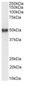
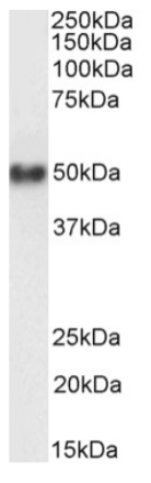
Western Blot using anti-CD38 antibody AT13/5 (Ab00289) Human spleen lysate samples (35µg protein in RIPA buffer) were resolved on a 10% SDS PAGE gel and blots probed with the chimeric rabbit version of AT13/5 (Ab00289-23.0) at 1 µg/ml before detection using an anti-rabbit secondary antibody. A primary incubation of 1h was used and protein was detected by chemiluminescence. The expected running size for CD38 is 34.3kDa, but this protein is glycosylated at several residues. Ab00289-23.0 successfully detected CD38 in human spleen lysate.
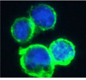
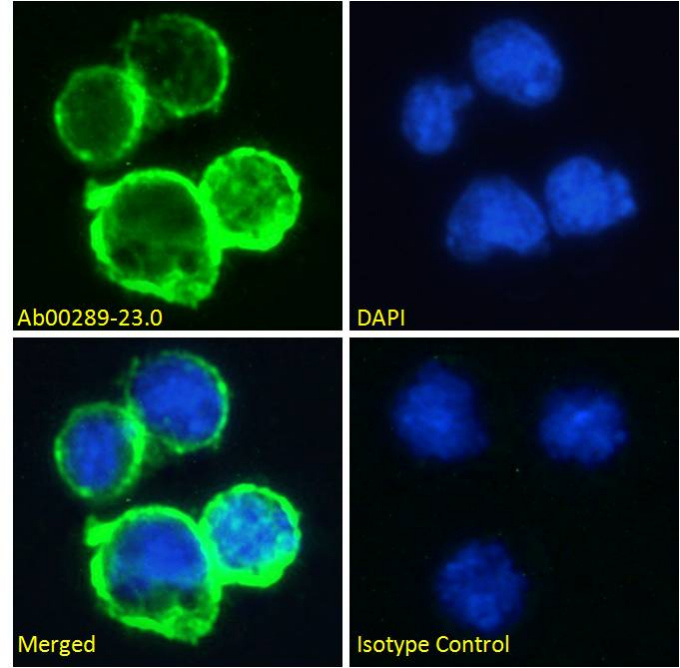
Immunofluorescence staining of fixed Daudi cells with anti-CD38 antibody AT13/5 (Ab00289) Immunofluorescence analysis of paraformaldehyde fixed Daudi cells on Shi-fix™ coverslips, permeabilized with 0.15% Triton and stained with the chimeric rabbit IgG version of AT13/5 (Ab00289-23.0) at 10 µg/ml for 1h followed by Alexa Fluor® 488 secondary antibody (1 µg/ml), showing membrane staining. The nuclear stain is DAPI (blue). Panels show from left-right, top-bottom Ab00289-23.0, DAPI, merged channels and an isotype control. The isotype control was stained with an anti-Fluorescein antibody (Ab102-23.0) followed by Alexa Fluor® 488 secondary antibody.