2
Products based on this clone:
- {{heading}}
Please Select
- Ab03205-23.0 Anti-E Cadherin [56-4]
- Mouse
- Rabbit IgG
- Purified
- In Stock
- Ab03205-1.1 Anti-E Cadherin [56-4]
- Mouse
- Mouse IgG1
- Purified
- In Stock
Loading...
 United Kingdom (UK)
Recombinant monoclonal antibody to E Cadherin. Manufactured using AbAb’s Recombinant Platform with variable regions (i.e. specificity) from the hybridoma 56-4.
UniProt Accession Number of Target Protein: P09803
Alternative Name(s) of Target: CD324; ARC-1; Cdh1; Cadherin-1; Cadherin E; E-Cadherin; Uvomorulin
Immunogen: The original antibody was generated by immunizing rabbits with purified extracellular domain of mouse E-cadherin.
Specificity: This antibody binds mouse E-cadherin. Cadherins are calcium-dependent cell adhesion proteins. It is localized on the surfaces of epithelial cells in regions of cell-cell contact known as adherens junctions. E-cadherins are reported to play an important role in cancer metastasis.
Application Notes: The binding of this antibody to mouse E-cadherin results in its activation. The initial binding characterization of this antibody to mouse E-cadherin was done using ELISA. This antibody was used to study the effect of activation of mouse E-cadherin on the growth and metastasis of a spontaneous breast cancer mouse model. Female MMTV-PyMT and control mice treated with 5 mg/kg of this antibody developed tumors in all 10 mammary glands. Also the number of metastasis nodules was significantly reduced with treatment with 56-4 activating antibody (PMID: 32127478; WO2020243616).
Antibody first published in:
Young Na et al. The functional activity of E-cadherin controls tumor cell metastasis at multiple steps. Proc Natl Acad Sci U S A. 2020 Mar 17;117(11):5931-5937. PMID:32127478
Note on publication:
The paper describes the use of activating antibodies to determine the role of E-cadherin in regulation of metastasis.
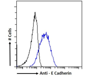
Flow cytometry using anti-E Cadherin antibody 56-4 (Ab03205). RAW 264.7 cells were stained with the anti-unknown specificity antibody (Ab00178-23.0; isotype control, black line) or the rabbit IgG version of 56-4 (Ab03205-23.0, blue line) at a dilution of 1:100 for 1h at RT. After washing, the bound antibody was detected using a goat anti-rabbit IgG AlexaFluor® 488 antibody at a dilution of 1:1000, and the cells were analyzed using a FACSCanto flow-cytometer.
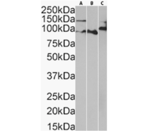
Western blot using anti-E Cadherin antibody 56-4 (Ab03205). Mouse lung (A) (0.01µg/ml), colon (B) (0.001µg/ml), and intestine (C) (0.1µg/ml) tissue lysates (35µg protein in RIPA buffer) were resolved on an SDS-PAGE gel, and blots were probed with the chimeric rabbit version of 56-4 (Ab03205-23.0) at the mentioned respective concentrations before detection using an anti-rabbit secondary antibody. A primary incubation of 1 hour was used, and protein was detected by chemiluminescence.

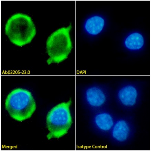
Immunofluorescence staining of RAW 264.7 cells with anti-E Cadherin antibody 56-4 (Ab03205). Immunofluorescence analysis of paraformaldehyde fixed RAW 264.7 cells on Shi-fix™ coverslips stained with the chimeric rabbit IgG version of 56-4 (Ab03205-23.0) (1:100 dilution) for 1h followed by Alexa Fluor® 488 secondary antibody (1:1000 dilution), showing membrane staining. The nuclear stain is DAPI (blue). Panels show, from left-right, top-bottom, Ab03205-23.0, DAPI, merged channels, and an isotype control. The isotype control was an unknown specificity antibody (Ab00178-23.0) followed by staining with Alexa Fluor® 488 secondary antibody.