2
Products based on this clone:
- {{heading}}
Please Select
- Ab01328-1.1 Anti-IgD [IgD26]
- Human
- Mouse IgG1
- Purified
- In Stock
- Ab01328-23.0 Anti-IgD [IgD26]
- Human
- Rabbit IgG
- Purified
- In Stock
Loading...
 United Kingdom (UK)
Recombinant monoclonal antibody to IgD. Manufactured using AbAb’s Recombinant Platform with variable regions (i.e. specificity) from the hybridoma IgD26.
UniProt Accession Number of Target Protein: P01880
Alternative Name(s) of Target: immunoglobulin D; immunoglobulin heavy constant delta; Igh-5; Ighd; immunoglobulin Delta
Immunogen: This antibody was raised by immunising mice with IgD purified from human myeloma serum.
Specificity: This antibody recognises human IgD which is a main antigen receptor on the surface of immature B-cells and small amount of it is secreted to the blood serum. On B-cells it takes part in the activation of these lymphocytes.
Application Notes: IgD26 may be used as a primary or secondary antibody. For instance, one group used anti-IgD antibody to label IgD in a cryostat tonsil section in the immunoperoxidase staining (Naiem et al., 1982).
Antibody first published in:
Taylor and Mason The immunohistological detection of intracellular immunoglobulin in formalin-paraffin sections from multiple myeloma and related conditions using the immunoperoxidase technique. Clin Exp Immunol. 1974 Nov;18(3):417-29. PMID:4219910
Note on publication:
This article describes the earliest application of this antibody present in the literature. The more detailed desription of the generation of such antibodies is described in the following paper: Naiem, M., Gerdes, J., Abdulaziz, Z., Sunderland, C. A., Allington, M. J., Stein, H., & Mason, D. Y. (1982). The value of immunohistological screening in the production of monoclonal antibodies. Journal of immunological methods, 50(2), 145-160.
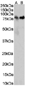
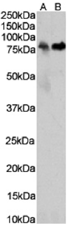
Western Blot using anti-IgD antibody IgD26 (Ab01328) human thymus (A) human plasma (B) tissue lysates (35µg protein in RIPA buffer) were resolved on a SDS PAGE gel and blots were probed with the chimeric rabbit version of IgD26 (Ab01328-23.0) at 0.03 µg/ml and 1 µg/ml, respectively, before detection using an anti-rabbit secondary antibody. A primary incubation of 1h was used and protein was detected by chemiluminescence.

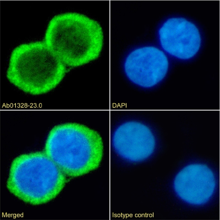
Immunofluorescence staining of fixed Daudi cells with anti-IgD antibody IgD26 (Ab01328) Immunofluorescence analysis of paraformaldehyde fixed Daudi cells on Shi-fix™ coverslips stained with the chimeric rabbit IgG version of IgD26 (Ab01328-23.0) at 10 µg/ml for 1h followed by Alexa Fluor® 488 secondary antibody (2 µg/ml), showing membrane staining. The nuclear stain is DAPI (blue). Panels show from left-right, top-bottom Ab01328-23.0, DAPI, merged channels and an isotype control. The isotype control was an unknown specificity antibody (Ab00178-1.1) followed by staining with Alexa Fluor® 488 secondary antibody.

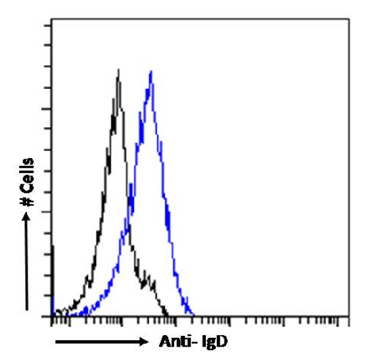
Flow cytometry using the Anti-IgD antibody IgD26 (Ab01328). Paraformaldehyde fixed Daudi cells were stained with anti-unknown specificity antibody (Ab00178-23.0; isotype control, black line) or the rabbit IgG version of IgD26 (Ab01328-23.0, blue line) at a dilution of 1:100 for 1h at RT. After washing, the bound antibody was detected using a goat anti-rabbit IgG AlexaFluor® 488 antibody at a dilution of 1:1000 and cells analyzed using a FACSCanto flow-cytometer.