2
Products based on this clone:
- {{heading}}
Please Select
- Ab01326-1.1 Anti-Lambda light chain [N10/2]
- Human
- Mouse IgG1
- Purified
- In Stock
- Ab01326-23.0 Anti-Lambda light chain [N10/2]
- Human
- Rabbit IgG
- Purified
- In Stock
Loading...
 United Kingdom (UK)
Recombinant monoclonal antibody to Lambda light chain. Manufactured using AbAb’s Recombinant Platform with variable regions (i.e. specificity) from the hybridoma N10/2.
UniProt Accession Number of Target Protein: P0DOY2
Alternative Name(s) of Target: IGL; immunoglobulin lambda light chain; lambda chain; lambda; λ immunoglobulin light chain; anti-λ
Immunogen: This antibody was raised by immunising BALB/c mice with purified human IgG.
Specificity: This antibody is specific for lambda light chains and does not show cross-reactivity with kappa light chains. It stains B cell follicles in human lymphoid tissues. Lambda light chain is one of the two types of light chains of mammalian immunoglobulins (one Ig has only one type of chains). Usually, the ratio of kappa to lambda is 70:30 but it might be disturbed in various B-cell conditions, such as myeloma.
Application Notes: N10/2 antibody may be utilised in the identification of certain pathologies (mainly associated with B-cells), such as leukemias, plasmacytomas, and some non-Hodgkin's lymphomas. For example, one group used this antibody during an immunohistochemical characterisation of human fetal bone sections engrafted with Waldenström macroglobulinemia cells (Tassone et al., 2005). Another group utilised N10/2 antibody to analyse the light chains expression in post-transplant lymphoproliferative disorders via immunohistochemistry on formalin-fixed, paraffin-embedded tissue samples (Capello et al., 2006).
Antibody first published in:
Mason et al. Double immunofluorescence labelling of routinely processed paraffin sections. J Pathol. 2000 Aug;191(4):452-61. PMID:10918222
Note on publication:
This article describes the earliest found application of this antibody where it was successfully utilised in immunohistochemical labelling.
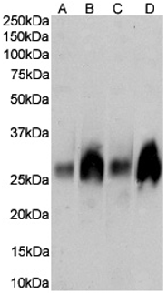
Western Blot using anti-Lambda light chain antibody N10/2 (Ab01326) Human spleen (A), human tonsil (B), human thymus (C) and human plasma (D) tissue lysates (35µg protein in RIPA buffer) were resolved on a SDS PAGE gel and blots were probed with the chimeric rabbit version of N10/2 (Ab01326-23.0) at 0.003 µg/ml, before detection using an anti-rabbit secondary antibody. A primary incubation of 1h was used and protein was detected by chemiluminescence.
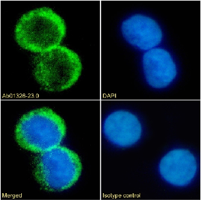
Immunofluorescence staining of fixed Daudi cells with anti-Lambda light chain antibody N10/2 (Ab01326) Immunofluorescence analysis of paraformaldehyde fixed Daudi cells on Shi-fix™ coverslips stained with the chimeric rabbit IgG version of N10/2 (Ab01326-23.0) at 10 µg/ml for 1h followed by Alexa Fluor® 488 secondary antibody (2 µg/ml), showing membrane staining. The nuclear stain is DAPI (blue). Panels show from left-right, top-bottom Ab01326-23.0, DAPI, merged channels and an isotype control. The isotype control was an unknown specificity antibody (Ab00178-1.1) followed by staining with Alexa Fluor® 488 secondary antibody.
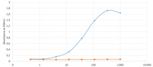
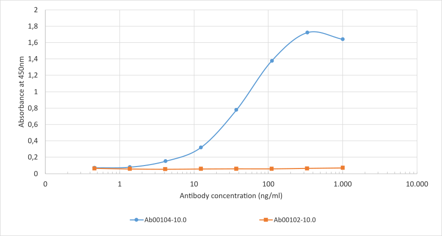
ELISA of anti-Lambda light chain antibody on a lambda chain antibody. Binding curves of the HRP-conjugated rabbit IgG chimeric version of the anti-Lambda light chain antibody N10/2 (Ab01326-23.0) to an ELISA plate coated with either a lambda chain antibody Ab00104-10.0 (B1-8); blue line) or a kappa light chain isotype control (Ab00102-10.0, anti-fluorescein; orange line) at a concentration of 2.5 µg/ml. A 3-fold serial dilution from 1000 ng/ml was tested in the experiment. Ab01326-23.0 bound only to the light chain antibody.
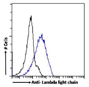
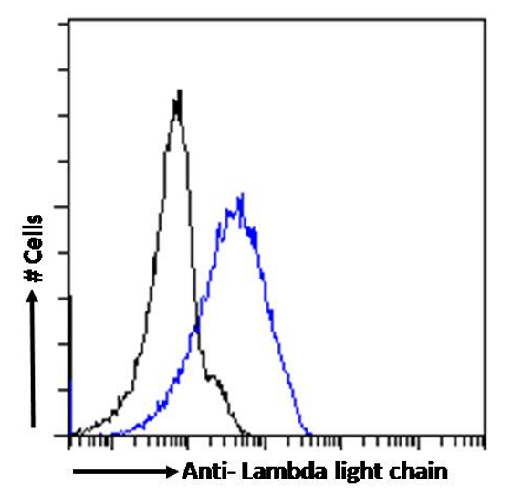
Flow cytometry using the Anti-Lambda light chain antibody N10/2 (Ab01326). Paraformaldehyde fixed Daudi cells were stained with anti-unknown specificity antibody (Ab00178-23.0; isotype control, black line) or the rabbit IgG version of N10/2 (Ab01326-23.0, blue line) at a dilution of 1:100 for 1h at RT. After washing, the bound antibody was detected using a goat anti-rabbit IgG AlexaFluor® 488 antibody at a dilution of 1:1000 and cells analyzed using a FACSCanto flow-cytometer.