2
Products based on this clone:
- {{heading}}
Please Select
- Ab01382-1.1 Anti-RAP [7F1]
- Human
- Mouse IgG1
- Purified
- In Stock
- Ab01382-23.0 Anti-RAP [7F1]
- Human
- Rabbit IgG
- Purified
- In Stock
Loading...
 United Kingdom (UK)
Recombinant monoclonal antibody to RAP. Manufactured using AbAb’s Recombinant Platform with variable regions (i.e. specificity) from the hybridoma 7F1.
Alternative Name(s) of Target: Receptor-associated protein; low density lipoprotein receptor-associated protein; 39-kDa receptor-associated protein
Immunogen: This antibody was raised by immunising mice with human placental RAP.
Specificity: This antibody is specific for human RAP. It does not cross-react with rabbit or murine RAP.
Application Notes: This antibody has been used to assess human RAP expression by RAP-transfected H4 cells, derived from human neuroglioma cells, in immunostaining analysis (Kinoshita et al, 2001). It has also been used in immunohistochemical analysis of paraffin-embedded human kidney tissue sections (Kounnas et al, 1992), and in ELISA analysis to detect RAP binding (Medh et al, 1995; Kounnas et al, 1993). Additionally, this antibody has been used to immunoprecipitate RAP, and in western blot analysis of purified B-amyloid protein and RAP mixtures (Kerr et al, 2010).
Antibody first published in:
Kounnas et al. The 39-kDa receptor-associated protein interacts with two members of the low density lipoprotein receptor family, alpha 2-macroglobulin receptor and glycoprotein 330. J Biol Chem. 1992 Oct 15;267(29):21162-6. PMID:1400426
Note on publication:
Describes the original generation of this antibody, and its use in immunohistochemistry.
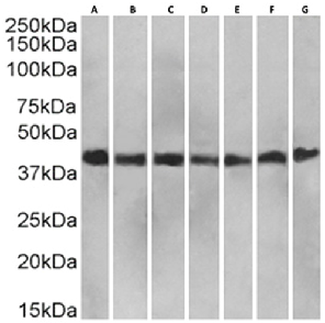
Western Blot using anti-RAP antibody (Ab01382) Staining in HeLa(A) (0.1ug/ml), A549 (B) (0.01ug/ml), HepG2 (C) (0.01ug/ml), HEK293 (D) (0.03ug/ml), MCF7 (E) (0.03ug/ml), Caco-2 (F) (0.001ug/ml) and U251 (G) (0.001ug/ml) cell lysates (35µg protein in RIPA buffer) were resolved on a SDS PAGE gel and blots were probed with the chimeric rabbit version of RAP (Ab01382-23.0) at 0.5 µg/ml, before detection using an anti-rabbit secondary antibody. A primary incubation of 1h was used and protein was detected by chemiluminescence.
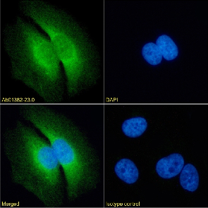
Immunofluorescence staining of fixed HeLa cells with anti-RAP antibody 7F1 (Ab01382) Immunofluorescence analysis of paraformaldehyde fixed HeLa cells on Shi-fix™ coverslips stained with the chimeric rabbit IgG version of 7F1 (Ab01382-23.0) at 10 µg/ml for 1h followed by Alexa Fluor® 488 secondary antibody (2 µg/ml), showing membrane staining. The nuclear stain is DAPI (blue). Panels show from left-right, top-bottom Ab01382-23.0, DAPI, merged channels and an isotype control. The isotype control was an unknown specificity antibody (Ab00178-23.0) followed by staining with Alexa Fluor® 488 secondary antibody.

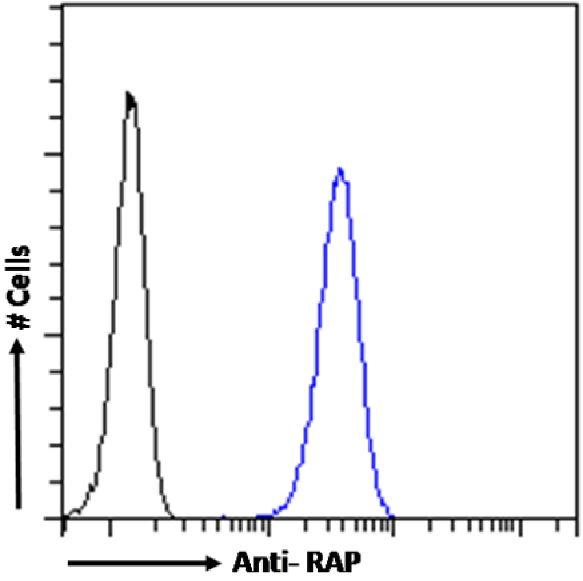
Flow cytometry using the anti-RAP antibody 7F1 (Ab01382). HeLa cells were fixed using 2% PFA and stained with anti-unknown specificity antibody (Ab00178-23.0; isotype control, black line) or the rabbit IgG1 version of 7F1 (Ab01382-23.0, blue line) at a dilution of 1:100 for 1h at RT. After washing, the bound antibody was detected using a goat anti-rabbit IgG AlexaFluor® 488 antibody at a dilution of 1:1000 and cells analyzed using a FACSCanto flow-cytometer.