2
Products based on this clone:
- {{heading}}
Please Select
- Ab01325-1.1 Anti-S100 [S1-61]
- Human
- Mouse IgG1
- Purified
- In Stock
- Ab01325-23.0 Anti-S100 [S1-61]
- Human
- Rabbit IgG
- Purified
- In Stock
Loading...
 United Kingdom (UK)
Recombinant monoclonal antibody to S100. Manufactured using AbAb’s Recombinant Platform with variable regions (i.e. specificity) from the hybridoma S1-61.
UniProt Accession Number of Target Protein: P23297
Alternative Name(s) of Target: S-100 protein; Protein S100-A1; S-100 protein alpha chain; S-100 protein subunit alpha; S100 calcium-binding protein A1; S-100
Immunogen: This antibody was raised by immunising BALB/c mice with S-100 protein, which was isolated from bovine brain andconjugated to BSA.
Specificity: This antibody is specific against an epitope located on the alpha-chain of S100. S100 is a protein antigen present at certain cell types and useful in anatomy and pathology. It is capable of binding calcium and is similar to calmodulin but its exact function is unclear.
Application Notes: This antibody is useful in determining the presence of S100 protein marker which might be used to characterise various normal and malignant cells. S1-61 antibody was initially used to immunohistochemically label cells in different tissues, such as nerve, striated muscle and melanoma, among others (Vanstapel et al., 1986). Another group utilised it to immunohistochemically identify strongly S100-positive extensions of central nervous system tissue in the area of dorsal root entry zones in patient samples with Friedreich ataxia (Koeppen et al., 2017). This antibody was shown to be also effective in detecting S100 on Western blot - one group analysed S100 expression in liver cells subjected to the activity of exosomes containing tumour-derived miRNA (Zeng et al., 2018).
Antibody first published in:
Vanstapel et al. Production of monoclonal antibodies directed against antigenic determinants common to the alpha- and beta-chain of bovine brain S-100 protein. Lab Invest. 1985 Feb;52(2):232-8. PMID:2578587
Note on publication:
This article describes generation and characterisation of S1-61 and (among other anti-S100 monoclonal antibodies).
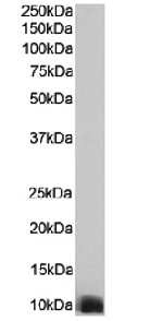
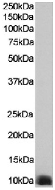
Western Blot using anti-S100 antibody S1-61 (Ab01325) Human heart tissue lysate (35µg protein in RIPA buffer) were resolved on a SDS PAGE gel and blots were probed with the chimeric rabbit version of S1-61 (Ab01325-23.0) at 0.00001 µg/ml, before detection using an anti-rabbit secondary antibody. A primary incubation of 1h was used and protein was detected by chemiluminescence.

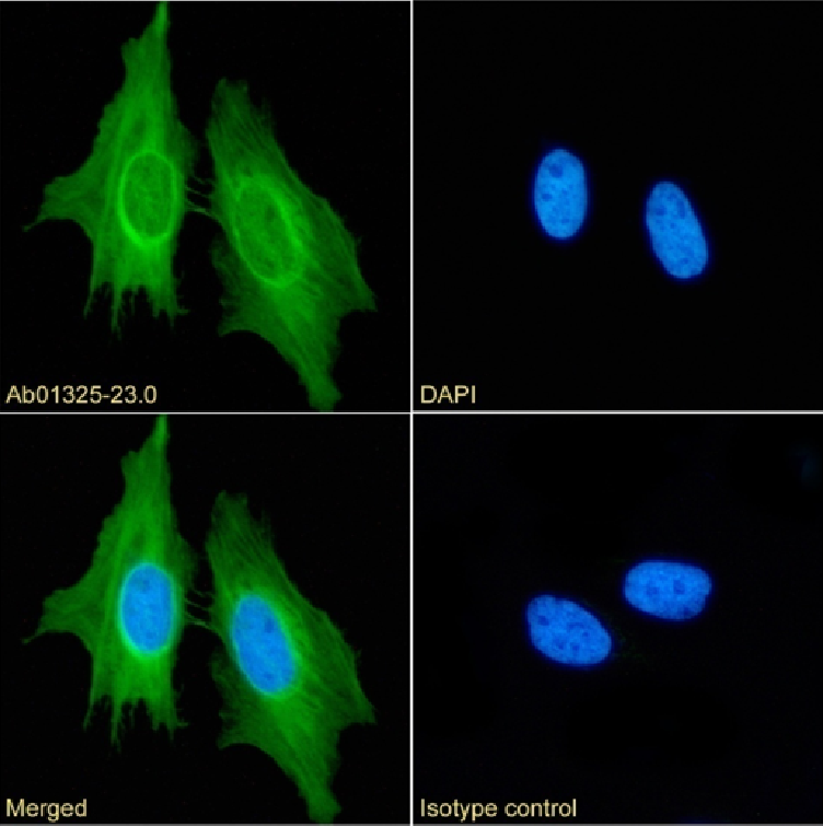
Immunofluorescence staining of fixed HeLa cells with anti-S100 antibody S1-61 (Ab01325) Immunofluorescence analysis of paraformaldehyde fixed HeLa cells on Shi-fix™ coverslips stained with the chimeric rabbit IgG version of S1-61 (Ab01325-23.0) at 10 µg/ml for 1h followed by Alexa Fluor® 488 secondary antibody (2 µg/ml), showing membrane staining. The nuclear stain is DAPI (blue). Panels show from left-right, top-bottom Ab01325-23.0, DAPI, merged channels and an isotype control. The isotype control was an unknown specificity antibody (Ab00178-1.1) followed by staining with Alexa Fluor® 488 secondary antibody.
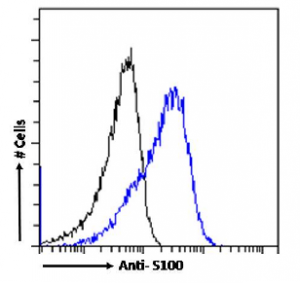
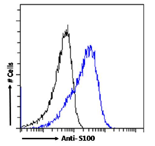
Flow cytometry using the Anti-S100 antibody S1-61 (Ab01325). Paraformaldehyde fixed HeLa cells were stained with anti-unknown specificity antibody (Ab00178-23.0; isotype control, black line) or the rabbit IgG version of S1-61 (Ab01325-23.0, blue line) at a dilution of 1:100 for 1h at RT. After washing, the bound antibody was detected using a goat anti-rabbit IgG AlexaFluor® 488 antibody at a dilution of 1:1000 and cells analyzed using a FACSCanto flow-cytometer.