- {{heading}}
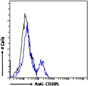
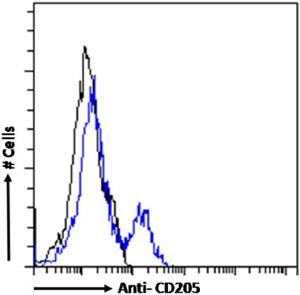
- Ab01616-21.0 Anti-CD205 [3B6]
- Chicken
- Mouse IgM
- Purified
- Ships in 4-5 weeks
- Ab01616-1.1 Anti-CD205 [3B6]
- Chicken
- Mouse IgG1
- Purified
- In Stock
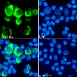
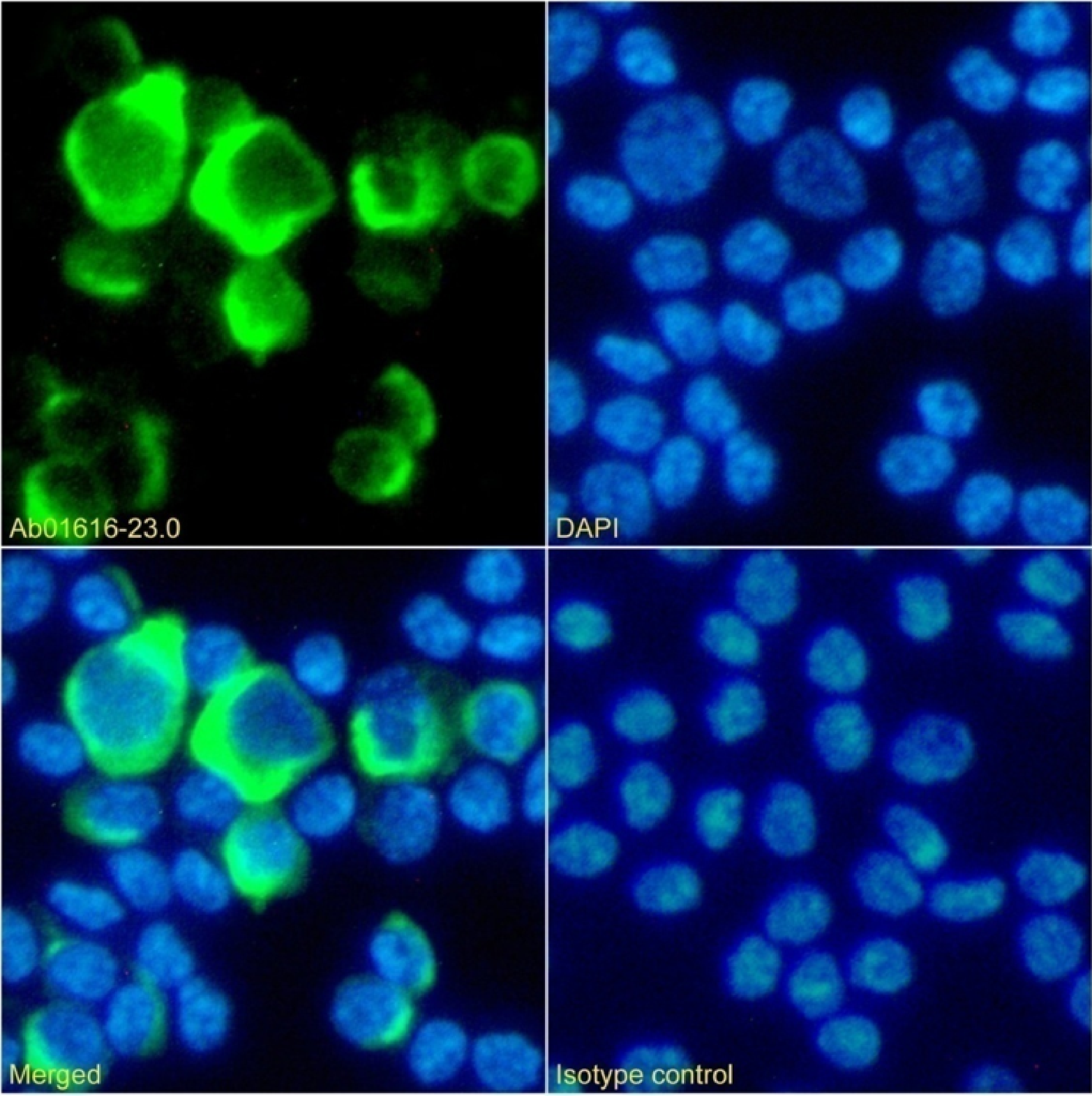
- Ab01616-23.0 Anti-CD205 [3B6]
- Chicken
- Rabbit IgG
- Purified
- In Stock
Recombinant monoclonal antibody to CD205. Manufactured using AbAb’s Recombinant Platform with variable regions (i.e. specificity) from the hybridoma 3B6.
Alternative Name(s) of Target: Lymphocyte antigen 75; LY75; Ly-75; C-type lectin domain family 13 member B; DEC-205; gp200-MR6
Immunogen: This antibody was raised by immunising mice against the recombinant C-type lectin domains 4-6 of the chicken CD205 protein.
Specificity: This antibody recognises chicken CD205 which is an endocytic receptor that is expressed at high levels by cortical thymic epithelial cells and by dendritic cell subsets, including the splenic CD8+ dendritic cell population. Antigen endocytosed via CD205 enters the MHC class I and MHC class II antigen presentation pathways and is subsequently presented to both CD4+ and CD8+ T cells (Shrimpton et al., 2009).
Application Notes: 3B6 antibody can be used to isolate chicken dendritic cells from peritoneal exudate cells; Vuong and colleagues (2019) did it using magnetic bead cell sorting. This allows performing further studies on this cell type in chicken. Furthermore, this antibody may also be used in the identification of the chicken dendtitic cells via immunofluorescence and cell sorting (Vuong et al., 2019).
Antibody first published in:
Vuong et al. CD205-positive, Sepharose-induced peritoneal exudate cells: a new resource for DC research in the chicken. Vet Res Commun. 2019 May;43(2):115-122. doi: 10.1007/s11259-019-09751-1. Epub 2019 Apr 16. PMID:30989431
Note on publication:
This article describes the generation and characterisation of 3B6 antibody.
Immunofluorescence staining of fixed chicken peripheral blood monocytes with anti-CD205 antibody 3B6 (Ab01616) Immunofluorescence analysis of paraformaldehyde fixed chicken peripheral blood monocytes on Shi-fix™ coverslips stained with the chimeric rabbit IgG version of 3B6 (Ab01616-23.0) at 10 µg/ml for 1h followed by Alexa Fluor® 488 secondary antibody (2 µg/ml), showing membrane staining. The nuclear stain is DAPI (blue). Panels show from left-right, top-bottom Ab01616-23.0, DAPI, merged channels and an isotype control. The isotype control was an unknown specificity antibody (Ab00178-23.0) followed by staining with Alexa Fluor® 488 secondary antibody.
Flow cytometry using the anti-CD205 antibody 3B6 (Ab01616). chicken peripheral blood monocytes were fixed using 2% PFA and stained with anti-unknown specificity antibody (Ab00178-23.0; isotype control, black line) or the rabbit IgG1 version of 3B6 (Ab01616-23.0, blue line) at a dilution of 1:100 for 1h at RT. After washing, the bound antibody was detected using a goat anti-rabbit IgG AlexaFluor® 488 antibody at a dilution of 1:1000 and cells analyzed using a FACSCanto flow-cytometer.