- {{heading}}
- Ab00242-3.0 Anti-CD7 [3A1E]
- Human
- Mouse IgG2b
- Purified
- In Stock
- Ab00242-23.0 Anti-CD7 [3A1E]
- Human
- Rabbit IgG
- Purified
- In Stock
- Ab00242-10.3 Anti-CD7 [3A1E]
- Human
- Human IgG1
- Fc Silent™
- Purified
- In Stock
Recombinant monoclonal antibody to CD7. Manufactured using AbAb’s Recombinant Platform with variable regions (i.e. specificity) from the hybridoma 3A1E.
UniProt Accession Number of Target Protein: P09564
Alternative Name(s) of Target: Leu9; T-cell antigen CD7; GP40; T-cell leukemia antigen; T-cell surface antigen Leu-9; TP41
Immunogen: This antibody was raised against CD7 expressing cells.
Specificity: Recognises human CD7.
Antibody first published in: Jansen et al. Successful treatment of human acute T-cell leukemia in SCID mice using the anti-CD7-deglycosylated ricin A-chain immunotoxin DA7. Cancer Res. 1992 Mar 1;52(5):1314-21. PMID:1371092


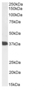
Western Blot using anti-CD7 antibody 3A1E (Ab00242) Jurkat cell lysate samples (35µg protein in RIPA buffer) were resolved on a 10% SDS PAGE gel and blots probed with the chimeric rabbit version of 3A1E (Ab00242-23.0) at 1 µg/ml before detection using an anti-rabbit secondary antibody. A primary incubation of 1h was used and protein was detected by chemiluminescence. The expected running size for unmodified CD7 is 25.4kDa, but this protein is glycosylated at several residues. Ab00242-23.0 successfully detected CD7 in Jurkat cell lysate.


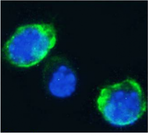
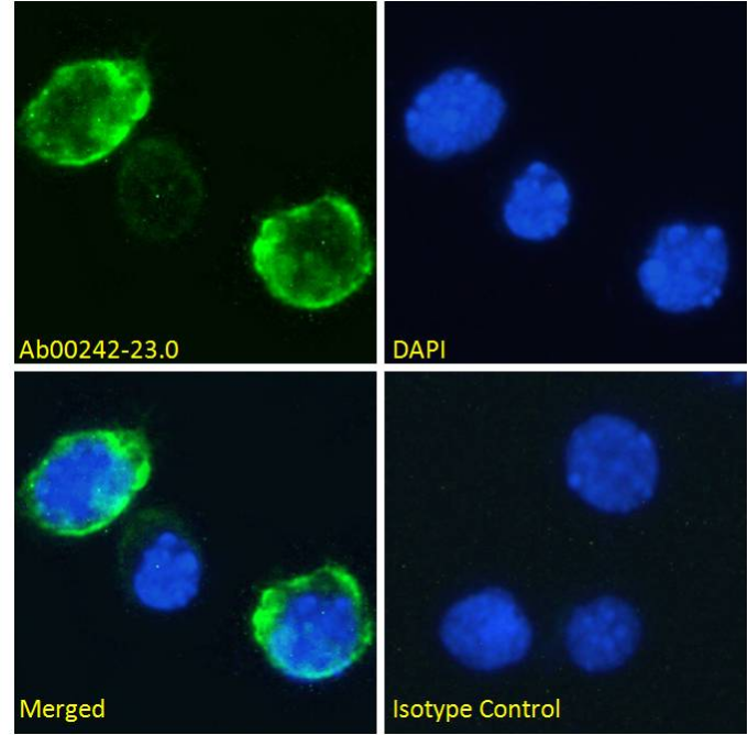
Immunofluorescence staining of fixed Jurkat cells with anti-CD7 antibody 3A1E (Ab00242) Immunofluorescence analysis of paraformaldehyde fixed Jurkat cells on Shi-fix™ coverslips, permeabilized with 0.15% Triton and stained with the chimeric rabbit IgG version of 3A1E (Ab00242-23.0) at 10 µg/ml for 1h followed by Alexa Fluor® 488 secondary antibody (1 µg/ml), showing membrane staining. The nuclear stain is DAPI (blue). Panels show from left-right, top-bottom Ab00242-23.0, DAPI, merged channels and an isotype control. The isotype control was stained with an anti-Fluorescein antibody (Ab102-23.0) followed by Alexa Fluor® 488 secondary antibody.