2
Products based on this clone:
- {{heading}}
Please Select
- Ab00329-23.0 Anti-UBE2I [SAIC-36A-9]
- Human
- Rabbit IgG
- Purified
- In Stock
- Ab00329-1.1 Anti-UBE2I [SAIC-36A-9]
- Human
- Mouse IgG1
- Purified
- In Stock
Loading...
 United Kingdom (UK)
Recombinant monoclonal antibody to UBE2I. Manufactured using AbAb’s Recombinant Platform with variable regions (i.e. specificity) from the B-cell cDNA library SAIC-36A-9.
UniProt Accession Number of Target Protein: P63279
Alternative Name(s) of Target: SUMO-conjugating enzyme UBC9; UBC9; UBCE9
Immunogen: The original antibody was raised against the KLH-conjugated peptide "DHPFGFVAVPTK", derived from the SUMO-conjugating enzyme UBC9.
Specificity: This antibody recognizes human UBE2I.
Application Notes: The original characterization data for this antibody can be found here.
Antibody first published in:
Schoenherr et al. Anti-peptide monoclonal antibodies generated for immuno-multiple reaction monitoring-mass spectrometry assays have a high probability of supporting Western blot and ELISA. Mol Cell Proteomics. 2015 Feb;14(2):382-98. doi: 10.1074/mcp.O114.043133 PMID:25512614
Note on publication:
The original publication describes the generation of a panel of recombinant antibodies using synthetic peptides of human proteins as well as their validation using ELISA and multiple reaction monitoring-mass spectrometry.
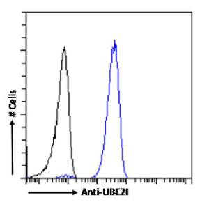
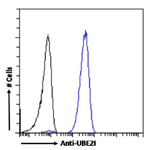
Flow cytometry using the Anti-UBE2I antibody SAIC-36A-9 (Ab00329). Paraformaldehyde fixed Jurkat cells permeabilized with 0.5% Triton were stained with anti-unknown specificity antibody (Ab00178-23.0; isotype control, black line) or the rabbit IgG version of SAIC-36A-9 (Ab00329-23.0, blue line) at a dilution of 1:100 for 1h at RT. After washing, the bound antibody was detected using a goat anti-rabbit IgG AlexaFluor® 488 antibody at a dilution of 1:1000 and cells analyzed using a FACSCanto flow-cytometer.

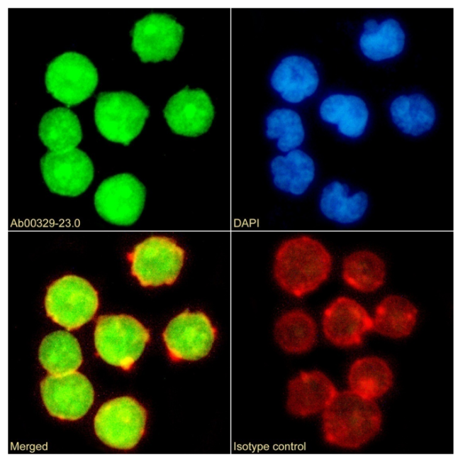
Immunofluorescence staining of Jurak cells with anti-UBE2I (Ab00329) SAIC-36A-9 Immunofluorescence analysis of paraformaldehyde fixed Jurak cells on Shi-fix™ cover-slips stained with the chimeric rabbit IgG version of SAIC-36A-9 (Ab00329-23.0) (1:100 dilution) for 1h followed by Alexa Fluor® 488 secondary antibody (1:1000 dilution), showing nuclear staining. Actin filaments were stained with phalloidin (red) and the nuclear stain is DAPI (blue). Panels show from left-right, top-bottom Ab00329-23.0, DAPI, merged channels and an isotype control. The isotype control was an unknown specificity antibody (Ab00178-23.0) followed by staining with Alexa Fluor® 488 secondary antibody.