 United Kingdom (UK)
 You have gone to great lengths to collect and suspend your cellular sample, and as antibody experts, we know the pain of ruined samples in the fixation and permeabilization steps of the flow cytometry process. Incidents like this can set back your project timeline and cause frustration in the lab. Common culprits of lab bench hiccups are the reagents you are using.
At Absolute Antibody, we are devoted to helping you run your experiments as efficiently and effectively as possible. Read on for how FIX&PERM® cell fixation and permeabilization buffers and recombinant antibodies can open new experimental possibilities and create reliably clean flow cytometry data trial after trial.
Gentle Cell Fixation and Permeabilization with FIX&PERM®
 Absolute Antibody is known for our engineered recombinant antibodies, but we’re excited to offer our clients easy access to other related reagents through our parent company Absolute Biotech, including the FIX&PERM® line of cell fixation and permeabilization buffers. This system utilizes two reagents and a simple protocol to prepare cells for flow cytometry. Reagent A gently fixes cells, while Reagent B permeabilizes cells. The FIX&PERM® formulation allows for simultaneous application of the permeabilization medium and antibodies conjugated with fluorescent compounds, enabling the staining of both intracellular and extracellular proteins.
FIX&PERM® buffers contain proven technology, so you know your cells are in good hands. Since the system was first characterized in 1994 by Knapp et al., FIX&PERM® has been independently validated multiple times (Groeneveld et al., 1996 and Lanza et al., 1997) and cited in thousands of studies for research on topics such as cancer biology, infectious disease monitoring, and more. Additional advantages include:
- Mild cell fixation, preserving their flow cytometric scatter characteristics
- Rapid technique – whole procedure can be carried out in less than one hour, ready for immediate analysis or storage for 24 hours
- Stringent QC procedures – the quality of each lot is confirmed using well-defined blood samples and subsequent comparison of scatter characteristics and immunostaining patterns, ensuring consistent and reliable results each time
- Stable – formulations do not contain volatile organic solvents, enabling storage at ambient temperature during shipment and in laboratories
- Flexible – can be used with most monoclonal antibody conjugates
- Simple protocol, starting with 50 µl of whole blood, bone marrow or mononuclear cell suspension (view protocol here)
- CE/IVD registered kits available for European customers in a clinical laboratory setting (contact us for further information)
Recombinant Antibodies for Flow Cytometry
Successful flow cytometry staining also requires using the right antibodies for the task, but not all antibodies are created equal. Mitigating nonspecific binding begins at the antibody selection stage, and recombinant monoclonal antibodies offer key advantages compared to traditional hybridoma-derived antibodies.
First, studies show that more than 30% of traditional hybridoma-derived monoclonal antibodies are not actually monoclonal. This means that the staining you achieve in your fixed and permeabilized cells may be nonspecific and cause background noise that alters your collected data. In contrast, recombinant antibody production starts at the sequence level, where the wholly defined antibody is expressed using synthetic genes in a well-characterized in vitro cell line. This animal-free platform guarantees the produced antibodies target the desired antigen without the usual risks of genetic drift and cell loss when compared to traditional hybridoma production methods.
Importantly, recombinant production also enables quick antibody engineering, enabling researchers to open experimental possibilities and refine existing protocols for flow cytometry applications. Our catalog of recombinant antibodies offers a variety of engineered options available off-the-shelf, including different isotypes, species and formats such as fragments. Reformatted antibodies can facilitate flow cytometry experiments by increasing compatibility with a secondary antibody, enabling easier co-labeling studies, and reducing non-specific binding. In particular, our Fc Silent™ format utilizes a point mutation to eliminate Fc binding and reduce background noise.
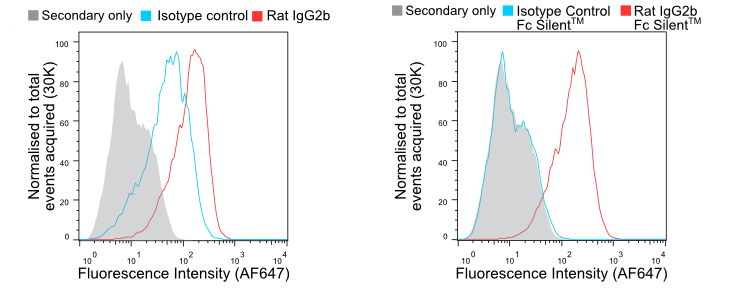
Figure 1. Flow cytometry of BMDMs stained with wild-type (A) and Fc Silent™ (B) anti-F4/80 (Ab00106-8.1 and Ab00106-8.4) and isotype control antibodies, followed by fluorescently conjugated goat anti-rat secondary antibody. Using Fc Silent™ abolishes non-specific FcγR driven staining, making data cleaner and more accurate.
Additionally, Absolute Antibody stores our recombinant antibodies in PBS solution without sodium azide, meaning nothing in our antibodies will adversely affect or degrade your cells. Antibodies can also be conjugated upon request with common fluorophores, such as FITC, PE, APC and FluoProbes 647h, as well as various tandem dyes.
Recombinant Antibodies in Action with Flow Cytometry
Recombinant antibodies from Absolute Antibody have been used to optimize existing protocols and create new protocols using flow cytometry methods. Below we describe some examples from recent literature.
This 2021 study by Wong et al. uses our human anti-CD98 antibody (Ab00361-10.0) as a reliable positive control for detecting CD98, a protein implicated in protective adaptive immunity in vertebrates, in flow cytometry assays to study the relationships between critically ill patients infected with COVID-19 and auto-reactive IgM antibodies. The methods for the above study are outlined in this article and are posited to be more cost-effective and faster than protein microarrays.
This 2019 study by Fultang et al. used the unconjugated research-grade biosimilar gemtuzumab (human IgG4; Ab00283-13.0) in a flow cytometry panel in order to test the cytotoxicity of the monoclonal therapeutic in comparison with its conjugated counterpart, gemtuzumab ozogamicin. In patient-derived, CD33+ myeloid-derived suppressor cells (MDSCs), both monoclonal antibody therapies were found to have cytotoxic effects on MDSCs. Recombinant antibody technology enables the effective analysis of monoclonal antibody therapies via flow cytometry, due in part to the 100% biological definition, so researchers know exactly the antibody sequences they are applying to their cells.
Another example of recombinant engineered antibodies from our catalog used for flow cytometry purposes appears in this 2018 paper published in Nature. Recombinant engineered anti-flavivirus group antigen/protein E antibodies (Ab00230-2.0) were incubated with ZIKV-infected cells fixed and permeabilized using FIX&PERM® for use in a flow cytometry assay to study the effect of semen on the Zika virus infection in cells from the anogenital tract. The study found that semen and seminal plasma inhibit ZIKV infection. A unique aspect of using recombinant antibodies from Absolute Antibody is the ability to choose from multiple formats. The format used in the aforementioned study was a mouse IgG2a, known for interacting relatively weakly with Fc receptors, potentially mitigating unwanted background. The Absolute Antibody catalog has a variety of formats for most of our antibody offerings.
High Quality Reagents for High Quality Data
With the amount of effort, time, and money that goes into sourcing, harvesting, and processing sample cells, it is crucial to treat them with the utmost respect throughout the experimental process. That means making the most of each cell with the protocols and materials you use. High-quality reagents, including FIX&PERM® cell buffers and recombinant engineered monoclonal antibodies, can ensure your cells stay intact throughout your experiments and keep unspecific binding to a minimum.
Try FIX&PERM® buffers through our Absolute Biotech sister brands Nordic-MUbio and Exalpha, and pair them with our engineered recombinant antibodies to make your flow cytometry as simple and effective as possible. Review a selection of our flow cytometry antibodies below. If we don’t have exactly what you need, or if you would like to explore your antibody options further, contact us or take a look at our custom service offerings!
Featured Recombinant Antibodies for Flow Cytometry
| Catalog Number | Clone Name | Antigen | Recombinant Formats Available |
|---|---|---|---|
| Ab01268 | RL388 | CD98 | Mouse IgG2a, Mouse IgG2a Fc Silent™, Rabbit IgG, Rat IgG2a |
| Ab00112 | UCHT1 | CD3 | Mouse IgG1, Mouse IgG1 Fc Silent™, Human IgG1, Human IgG1 Fc Silent™, Rabbit IgG |
| Ab00606 | RFB-4 | CD22 | Mouse IgG1, Rabbit IgG |
| Ab02772 | 4C5 | Lactoferrin | Mouse IgG1, Rabbit IgG, Human IgG1, Human IgG1 Fc Silent™ |
| Ab02769 | LZ-2 | Lysozyme | Mouse IgG1, Rabbit IgG, Human IgG1, Human IgG1 Fc Silent™ |
| Ab00613 | FMC63 | CD19 | Mouse IgG2a, Human Fab fragment, Mouse IgG2a Fc Silent)tm), Human IgG1, Rabbit IgG, Mouse Fab fragment, scFv fragment (His) |
| Ab00103 | 4D5-8 (trastuzumab) | erbB-2 (Her-2/neu) | Human IgG1 |
| Ab00126 | 10F381 (rituximab) | CD20 | Human IgG1, Human IgG1 Fc Silent™, Rabbit IgG, Mouse IgG2a, Cynomolgus monkey IgG1, Rhesus macaque IgG1, Mouse IgA, Mouse IgM, Human IgA1, Human IgM, Human IgG1 STR Fc-Silenced |
| Ab01258 | 1B4 | TIGIT | Mouse IgG1 |
| Ab00124 | Campath-1H | CD52 | Human IgG1 |
| Ab00279 | C225 (Cetuximab) | EGFR | Human IgG1 |
| Ab00449 | M200 (Volociximab) | alpha 5 beta 1 Integrin | Rabbit IgG |
| Ab00813 | RMP1-14 | PD-1 | Mouse IgG2a, Mouse IgG2a Fc Silent™, Rat IgG2a, Rabbit IgG, Mouse IgG1-D265A Fc Silenced, Mouse IgG1, Mouse bispecific (anti-mPD-L1), Mouse bispecific (anti-mCD47), Mouse IgG2c Fc Silent™, Mouse Fab fragment His-Tagged |
| Ab00779 | ZKA64 | Envelope Protein | Human IgG1, Human Fab fragment His-Tagged, Rabbit IgG, Human IgM, Mouse IgG2a, Mouse IGG2a Fc Silent™, Human IgG1-LALA Fc Silenced, Ferret IgG1, Ferret IgM, Ferret IgA |
| Ab00715 | bevacizumab | VEGF | Human IgG1, Human IgG1 Fc Silent™, Rabbit IgG, Mouse IgG2a, Mouse IgM, Human IgM, Human Fab fragment His-Tagged |
| Ab01419 | 10F.9G2 | PD-L1 | Rat IgG2b, Rat IgG2b Fc Silent™, Mouse IgG2b, Mouse IgG2b Fc Silent™, Rabbit IgG, Mouse Fab fragment His-Tagged, Mouse bispecific (anti-mOX40), Mouse IgG2a mCD3e KIH bispecific Fc Silent™, Mouse IgG2a mCD28 KIH bispecific Fc Silent™ |
| Ab02019 | CV30 | Spike Protein (RBD) | Human IgG2, Human IgG3, Human Fab fragment His-Tagged, Human F(ab)2 His-Tagged, Mouse Fab fragment His-Tagged, Human IgG1, Human IgG1 Fc Silent™, Human IgM, Rabbit IgG, Mouse IgM, Human IgA1, Mouse IgG1, Cat IgM, Cat IgG1b, Ferret IgG1, Ferret IgM, Ferret IgA, Human IgA2, Human IgG4-S228P, Human IgE, Human IgA (J-Chain) |
| Ab00187 | daclizumab | IL-2R | Human IgG1, Human IgG1 Fc Silent™, Rabbit IgG, Mouse IgG1, Mouse IgG2a, Cynomolgus monkey IgG1, Rhesus macaque IgG1, Mouse IgG2a Fc Silent™ |
| Ab00728 | hL22 (epratuzumab) | CD22 | Human IgG1, Human IgG1 Fc Silent™, Rabbit IgG, Cynomolgus monkey IgG1, Cynomolgus monkey IgG4, Rhesus macaque IgG1, Rhesus macaque IgG4 |
| Ab00146 | cA2 | TNF alpha | Human IgG1, Human IgG1 Fc Silent™, Rabbit IgG, Mouse IgG2a, Mouse IgA, Mouse IgM, Human IgA1, Human IgM |
| Ab00221 | K20 | CD29 | Mouse IgG2a, Rabbit IgG |
| Ab01489 | 6D5 | CD19 | Rat IgG2a, Mouse IgG2a, Mouse IgG2a Fc Silent™, Rabbit IgG, Mouse IgG2a mCD3e KIH bispecific Fc Silent™ |
| Ab00447 | hu5c8 (ruplizumab) | CD40L | Human IgG1, Mouse IgG1, Mouse IgG1 Fc Silent™, Rabbit IgG, Mouse IgG2a, Human IgG1 Fc Silent™ |
| Ab00450 | CE9.1 (clenoliximab) | CD4 | Human IgG4, Mouse IgG1, Mouse IgG1 Fc Silent™, Rabbit IgG |
| Ab01107 | PC-61.5.3 | CD25 | Rat IgG2a, Rat IgG2a Fc Silent™, Mouse IgG2a, Mouse IgG2a Fc Silent™, Mouse IgG1-D265A Fc Silenced, Rabbit IgG, Mouse Fab fragment His-Tagged, Rat IgG1 |
| Ab00282 | 7E3 (abciximab) | CD41 | Human IgG1, Rabbit IgG, Human IgG1 Fc Silent™, Mouse IgG2a, Human Fab fragment His-Tagged, Human IgG4 |
| Ab01420 | 13F3 | VISTA | Hamster IgG, Mouse IgG2b, Mouse IgG2b Fc Silent™, Mouse IgG2-D265A Fc Silenced, Rabbit IgG, Mouse Fab fragment His-Tagged, Mouse IgG2a |
Latest News
Upcoming Events
Please join us at the following conferences and events. Stop by our booth, or get in touch to arrange a meeting.
See All Dates