 United Kingdom (UK)
 Immunotherapy has emerged as a compelling therapeutic approach for the effective treatment of various tumors. Immunotherapy is based on strengthening the immune system in order to recognize and attack tumor cells (Tan et al., 2020). Members of the B7 family, such as programmed death ligand 1 (PD-L1, B7-H1, CD274) and PD-L2 (B7-DC, CD273), have been shown to be important for regulating the immune response, therefore playing a significant role in the immunosuppression of tumors (Ni et al., 2017).
Monoclonal antibodies against the two programmed ligands, PD-L1 and PD-L2, are valuable tools to sharpen our understanding of the PD-1:PD-L1/2 pathway. Absolute Antibody offers a large selection of anti-PD-L1 and anti-PD-L2 antibodies (table below), including the anti-mouse PD-L1 clone as an IgG2b and IgG2b Fc Silent™, the anti-PD-L2 clone TY25 as a mouse IgG2a Fc Silent™, and various anti-PD-1 antibodies.
PD-L1 and PD-L2 are type I transmembrane glycoproteins, containing IgC and IgV domains. PD-L1 is expressed by T cells, B cells, NK cells, dendritic cells, macrophages, MDSCs, and many other cell types such as epithelial and endothelial cells, whereas PD-L2 is primarily expressed on professional antigen presenting cells including dendritic cells (DCs) and macrophages (Hudson et al., 2020).
The two programmed death ligands, PD-L1 and PD-L2, respectively interact with PD-1, forming immune checkpoint regulators that suppress immune responses and promote self-tolerance (Latchman et al., 2001). However, the two ligands show distinct affinity, molecular mechanisms of interaction, and functional differences with PD-1. Further, they differ in specificity, since PD-L1 but not PD-L2 can bind also to CD80 (Ghiotto et al., 2010). The PD-1:PD-L1/2 checkpoints are often exploited by malignant tumors to evade immune surveillance. Therefore, targeting PD-L1 and PD-L2 with inhibitory antibodies results in successful enhancement of T cell immunity against tumors (Bardhan et al., 2016). However, further research is needed to shine light on the therapeutic mechanisms of action of the two ligands (Lee et al., 2019).
Recombinant Engineered Antibodies
To bridge the gaps in understanding the therapeutic potential of PD-1’s ligands, Absolute Antibody recombinantly produces several anti-PD-L1 and anti-PD-L2 antibodies targeting PD-L1 in a variety of formats and species. A crucial step on the preclinical pathway is using live mouse models to study the in vivo effects of anti-PD-L1/L2 antibodies as therapeutic options. To address this need, our VivopureX™ line offers antibodies with mouse-anti-mouse backbones, produced with industry-leading purity standards to reduce immunogenicity and maintain therapeutic performance in vivo over time.
The VivopureX™ line includes anti-PD-L1 and anti-PD-L2 clones built specifically for optimized in vivo use. The anti-PD-L1 clone 10F.9G2 is species-swapped from the original rat IgG2b to mouse IgG2b for better therapeutic outcomes and cleaner data, particularly for blocking applications. Additionally, the anti-PD-L1 mouse-anti-mouse format is offered with our Fc Silent™ modification to abrogate Fc region binding for tailored effector function. We confirmed specificity of this clone using ELISA analysis (Fig. 1, Ab01419, blue line).
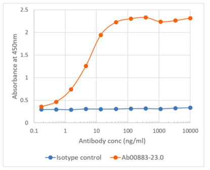
We also have the anti-PD-L2 clone TY25 available in mouse IgG2a Fc Silent™ format, species-swapped and engineered from its original rat IgG2a format for better performance in blocking applications. See Figure 2 for ELISA analysis of this clone’s confirmed specificity.
Our VivopureX™ antibodies empower in vivo studies, especially in the field of cancer research, with clinically relevant antibody formats. Further, our VivopureX™ catalog includes antibodies against other members of the B7 family, such as our anti-mouse CD80 (RM80 Fc Silent™) and VISTA (13F3 Fc Silent™) clones.
Please contact us if you are interested in anti-PD-L1 and anti-PD-L2 antibodies, their applications, or their availability in different formats. The full list of our anti-PD-L1 and anti-PD-L2 clones, antigens, reported applications, and formats currently available on the website are depicted in the table below. Follow the product links to view available characterization data.
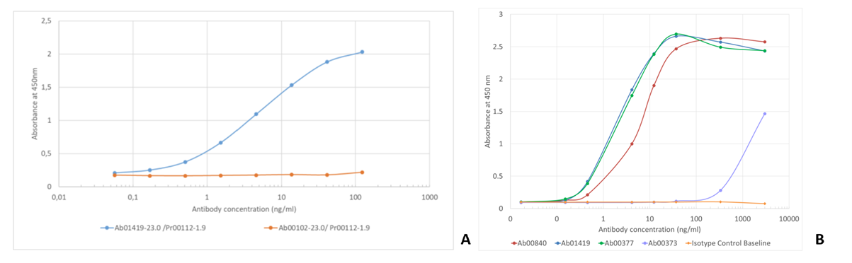
Fig. 1. (A) ELISA using 10F.9G2 (Ab01419, blue line) binding to recombinant mouse PD-L1 Fc fusion protein and (B) ELISA using 10F.9G2 (blue line) binding to recombinant mouse PD-L1 Fc fusion protein.
| Catalog Number | Clone Name | Antigen | Applications | Recombinant Formats Available |
|---|---|---|---|---|
| Ab00373 | YDC 127.1.1 | PD-L1 | FC | Rabbit IgG Mouse IgG1-D265A Mouse IgG2a Fc Silent™ Mouse Bispecific, anti-mCD47 Rat IgG2a Rat IgG2a Fc Silent™ |
| Ab00377 | alphaPD-L1 | PD-L1 | ELISA; Blocking | Rabbit IgG Mouse IgG1 Mouse IgG1 Fc Silent™ Mouse IgG1-D265A Mouse F(ab)2 |
| Ab00840 | B3 | PD-L1 | block; in vivo; FC; ELISA; functional assay | Rabbit IgG-Fc fusion |
| Ab01419 | 10F.9G2 | PD-L1 | ELISA; FC; IHC; blocking | Rabbit IgG Mouse IgG2b Mouse IgG2b Fc Silent™ Mouse Fab fragment Mouse Bispecific, anti-mOX40 Mouse IgG2a mCD3e KIH bispecific Mouse IgG2a mCD28 KIH bispecific Rat IgG2b Rat IgG2b Fc Silent™ |
| Ab02248 | JC071 | PD-L1 | FC, WB, ELISA, Block | Rabbit IgG Mouse IgG1 Dog IgG2 (IgG-B) Dog IgG2 (IgG-B) Fc Silent™ Dog IgG4 (IgG-D) |
| Ab02428 | 332M7 | PD-L1 | FC; block | Rabbit IgG Human IgG1 Human IgG1 Fc Silent™ Mouse IgG2b Mouse IgG2b Fc Silent™ |
| Ab02429 | B7HC0013 | PD-L1 | ELISA; IHC | Rabbit IgG Human IgG1 Human IgG1 Fc Silent™ Mouse IgG1 |
| Ab02430 | XGMHc25.1 | PD-L1 | FC; IHC | Rabbit IgG Human IgG1 Human IgG1 Fc Silent™ Mouse IgG2b Mouse IgG2b Fc Silent™ |
| Ab02431 | AB1 | PD-L1 | ELISA; FC | Rabbit IgG Human IgG1 Human IgG1 Fc Silent™ Mouse IgG2b Mouse IgG2b Fc Silent™ |
| Ab03442 | 5H1 | PD-L1 | FC; ELISA | Rabbit IgG scFv fragment (His) |
| Ab03452 | 29E.2A3 | PD-L1 | FC; blocking; IHC | Rabbit IgG Human IgG1 Human IgG1 Fc Silent™ Mouse IgG2b Mouse IgG2b Fc Silent™ |
| Ab03505 | mAb #18 (PD-L1.A) | PD-L1 | Inhibition; SPR; WB; in vivo | Rabbit IgG Human IgG1 Human IgG1 Fc Silent™ Mouse IgG1 |
| Ab03773 | 14-02-3 | PD-L1 | FC | |
| Ab03784 | BT613 | PD-L1 | SPR; blocking; in vitro; inhibition; in vivo | Rabbit IgG Human IgG1 Human IgG1 Fc Silent™ Mouse IgG1 |
| Ab03785 | CA782 | PD-L1 | ELISA; SPR | Rabbit IgG Human IgG1 Human IgG1 Fc Silent™ Mouse IgG1 |
| Ab00852 | Z64P2D3*H4 | PD-L2 | ELISA; WB; IHC | Rabbit IgG Human IgG1 Human IgG1 Fc Silent™ Human IgG4-S228P Mouse IgG1 |
| Ab00883 | TY25 | PD-L2 | FC; IP; WB; Block | Rabbit IgG Mouse IgG2a Mouse IgG2a Fc Silent™ Mouse IgG1-D265A Rat IgG2a |
| Ab01479 | 366C.9E5 | PD-L2 | IHC | Rabbit IgG Human IgG1 Human IgG1 Fc Silent™ Human IgG4-S228P Mouse IgG1 |
| Ab02439 | 24F.10C12 | PD-L2 | ELISA; FC; IHC; Block | Rabbit IgG Human IgG1 Human IgG1 Fc Silent™ Human IgG4-S228P Mouse IgG1 |
| Ab03771 | HZ-D-Na-96-1 | PD-L2 | FC; block | |
| Ab03772 | HZ-D-Ye-29-3 | PD-L2 | FC; block | Rabbit IgG Human IgG1 Human IgG1 Fc Silent™ Mouse IgG2a Mouse IgG2a Fc Silent™ |
Article compiled by Dr. Lucia Lupica-Spagnolo, Absolute Antibody Product Development Manager
Latest News
Upcoming Events
Please join us at the following conferences and events. Stop by our booth, or get in touch to arrange a meeting.
See All Dates